About

Tag: worms

  • Would you still love me if I were a worm?

    Would you still love me if I were a worm?

    By Michelle Cheng

    ~12 minutes


    How Scientists are Using Worms to Learn About Humans

    Worms and humans could not possibly be any more different. And yet, scientists have been studying C. elegans (caenorhabditis elegans) to learn more about the human body over 70 years. These unassuming worms have aided in groundbreaking findings in medicine for human diseases such as Alzheimer’s, AIDS, and stroke.

    What makes C. elegans so valuable is not its complexity, but rather its simplicity. Because so many of its biological pathways are conserved in humans, this worm provides a uniquewindow into the fundamental processes of life, including cell division, gene regulation, neural signaling, and aging. With a transparent body, rapid life cycle, and a genetic makeup that mirrors much of our own, C. elegans has become an essential organism in modern biomedical research. Understanding how scientists use these worms can help us appreciate not just what we’ve already learned, but also the vast potential that still lies ahead.

    What is C. elegans?

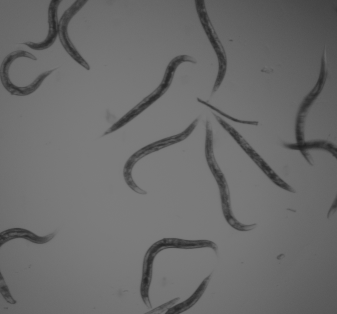
    C. elegans is a free-living nematode that has become one of the most important model organisms in research. It measures approximately one millimeter in length and naturally lives in temperate soil environments, where it feeds on bacteria like e. coli. It is non-parasitic and exists in two sexes: hermaphrodites, which are capable of self-reproduction, and males, which occur at a less than 0.1% chance under normal conditions. The hermaphroditic reproductive mode allows for the maintenance of isogenic populations, which is advantageous for genetic studies.

    Anatomy of an adult C. elegans hermaphrodite / EnVivo Biosystems ©

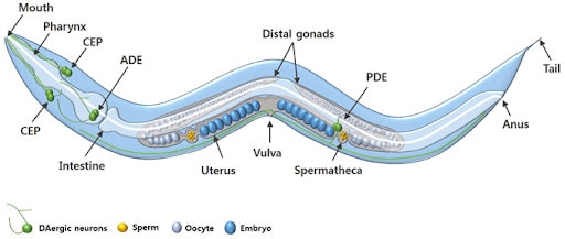
    The adult C. elegans hermaphrodite has exactly 959 somatic cells while the adult male C. elegans has exactly 1,031 somatic cells. The worm’s relatively simple anatomy includes muscles, a nervous system, a digestive system, a reproductive system, and an excretory system. The organism develops through four larval stages before reaching adulthood, with a complete lifecycle taking just two to three weeks under laboratory conditions.

    The life cycle of C. elegans / National Institute of Health ©

    Genetically, C. elegans has a compact genome consisting of about 100 million base pairs across six chromosomes. It was the first multicellular organism to have its entire genome sequenced in 1998 in a project led by John Sulston and Bob Waterstons. Its genome is highly amenable to manipulation using a variety of modern techniques.

    Why do scientists study C. elegans specifically?

    First introduced into studies by Sydney Brenner in the 1960s to study neurological development and the nervous system, the nematode proved itself in the lab with its unique combination of genetic, anatomical, and practical features that make it exceptionally suitable for biomedical research. 

    Remarkably, around 60-70% of human disease-associated genes have counterparts in the C. elegans genome, making it an incredibly valuable model for studying human biology. Many genes responsible for critical cellular functions are evolutionarily conserved between worms and humans. Therefore, scientists can manipulate the function of these genes in C. elegans to study their roles in disease without the complexity or ethical challenges of working with human subjects or higher animals like mice or primates. 

    Analogous counterparts of the human nervous systems in C. elegans / Taylor and Francis Online ©

    Adult hermaphrodites’ cells, which remain the same in every single  worm, each of which has been identified and mapped, allowing for detailed tracking of development, differentiation, and cellular processes. Its transparent body enables real-time visualization of internal structures, including neurons, muscles, reproductive organs, and digestive tissues. The worm, which has a simple nervous system of only 302 cells, is one of the only organisms where every neural connection is known. Additionally, C. elegans has a short life cycle of two to three weeks and is easy to culture in large numbers, making it especially convenient for developmental and aging studies.

    How do scientists modify C. elegans in experiments?

    Scientists modify and study C. elegans using four primary methods: RNA interference (RNAi), CRISPR-Cas9 genome editing, transgenic techniques, and drug screening.

    Different modes of administration of dsRNAs for RNA interference / Biomed Central ©

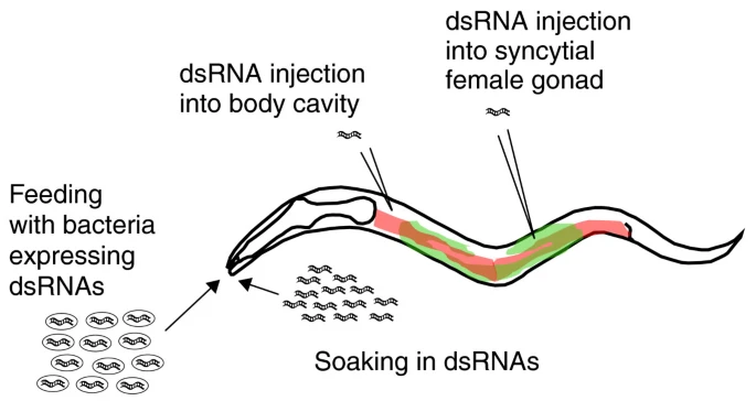
    One of the most widely used techniques for modifying gene expression in C. elegans is RNA interference (RNAi). This method allows scientists to silence specific genes to observe the effects of their absence. In C. elegans RNAi can be easily administered by feeding worms with genetically engineered E. coli bacteria that produce double-stranded RNA (dsRNA) matching the gene of interest. Once ingested, the dsRNA activates the worm’s endogenous RNAi pathway, leading to the degradation of the corresponding messaging RNA and a reduction or elimination of the target protein. This method is highly efficient, non-invasive, and relatively easy to perform, making it ideal for large-scale genetic screens. Researchers can identify genes involved in key processes such as embryonic development, aging, metabolism, and neurodegeneration.

    The CRISPR-Cas9 system has revolutionized genetic research in C. elegans by enabling precise, targeted alterations to the genome. Scientists introduce a complex composed of the Cas9 enzyme and a guide RNA (gRNA) into the worm, which directs the Cas9 to a specific DNA sequence. Once there, Cas9 introduces a double-strand break in the DNA. The cell’s natural repair mechanisms then fix the break, and researchers can insert, delete, or replace specific DNA sequences. In C. elegans, CRISPR can create mutants mimicking  human disease alleles or study regulatory elements of genes. This method provides a level of control that surpasses RNAi, as it allows for permanent and heritable genetic modifications. Scientists often inject the CRISPR-Cas9 components directly into the gonads of adult hermaphrodites, ensuring that the genetic changes are passed onto the offspring.

    Image of the pharynx of C. elegans expressing GFP / Leica Microsystems ©

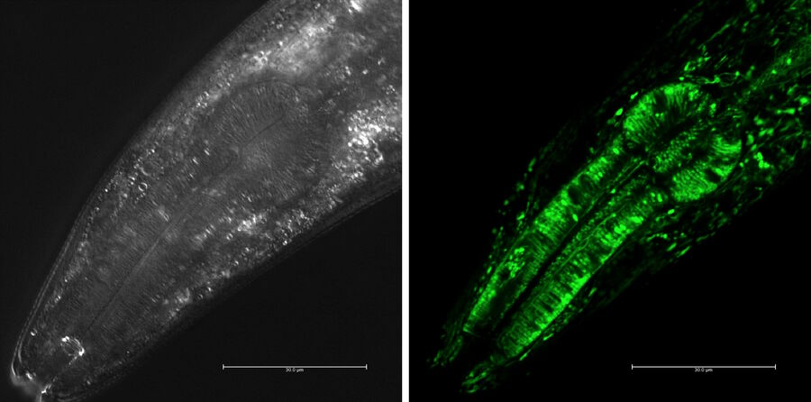
    Transgenic techniques in C. elegans insert foreign DNA into the worm’s genome to monitor gene expression, trace cell lineages, or study protein localization. One common approach is to fuse a gene of interest to a reporter gene such as green fluorescent protein (GFP). When this gene is expressed, the fluorescent tag can be visualized in living worms using fluorescence microscopy. This allows researchers to observe where and when specific genes are active, how proteins move within the cells, and how cells interact during development or disease progression. Transgenes are typically introduced via microinjection into the syncytial gonads of adult worms, leading to the formation of extrachromosomal arrays inherited by the next generation. Stable lines can also be created through CRISPR or chemical integration methods. These visual tools are particularly powerful due to the worm’s transparent body, which makes it possible to track fluorescent signals in real time throughout the entire organism.

    C. elegans is an excellent system for drug screening and environmental toxicology due to its small size, short lifespan, and genetic tractability. Researchers can test the effects of thousands of compounds quickly and cost-effectively. In these experiments, worms are exposed to chemical agents in liquid or on agar plates, and their survival, movement, reproduction, or specific cellular markers are measured to assess the biological impact. Using genetically modified strains that mimic human disease pathways, scientists can screen for drugs that alleviate symptoms or restore normal function. These tests provide an efficient first step in drug development, singling out promising candidates before moving onto mammalian models.

    The cell lineage and the programmed cell death in C. elegans / Nobel Prize in Physiology or Medicine 2002

    One of the most groundbreaking discoveries made using C. elegans was the genetic basis of programmed cell death, or apoptosis, a critical process in both development and disease. The research was led by Dr. H. Robert Horvitz at the Massachusetts Institute of Technology. Horvitz and his colleagues began studying cell death in C. elegans in the 1980s by tracing the fate of every cell in the worm’s body during development. They discovered that exactly 131 cells always die in the developing hermaphrodite and that this process was genetically controlled. Through genetic screening, Horvitz identified three core genes that regulated apoptosis: ced-3, ced-4, and ced-9. By inducing mutations in these genes, the researchers could either prevent or accelerate cell death in the worm. This revealed that cell death is not a passive consequence of damage, but rather an active, genetically programmed event. The mammalian counterparts of these genes, like caspases and BCL-2, were later discovered to play central roles in cancer, autoimmune diseases, and neurodegeneration, making this research foundational to modern medicine. Horvitz was awarded the 2002 Nobel Prize in Physiology or Medicine for his work along with Sydney Brenner and John Sulston.

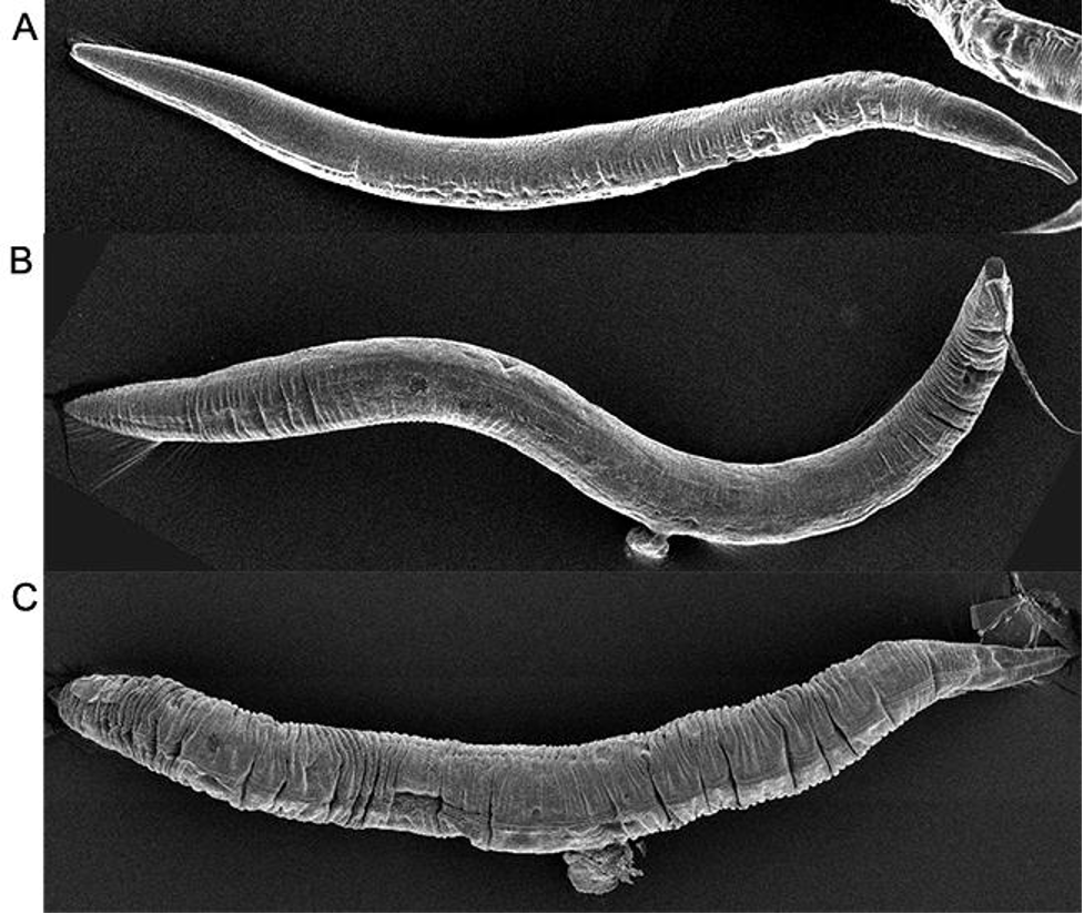
    In addition, C. elegans has contributed to our understanding of neurodegenerative diseases such as Alzheimer’s. One major study was led by Dr. Christopher Link at the University of Colorado in the late 1990s. Link developed a transgenic C. elegans strain that expressed the human β-amyloid (Aβ) peptide in muscle cells. This is the same peptide that forms toxic plaques in the brains of Alzheimer’s patients. In the study, the researchers observed that worms expressing Aβ developed progressive paralysis as they aged, mimicking aspects of human Alzheimer’s pathology. They then used this model to screen for genetic mutations and chemical compounds that could suppress the toxic effects of Aβ. Their work identified several genes involved in protein folding and stress response that modified Aβ toxicity. This demonstrated that C. elegans could be used as a fast and cost-effective in vivo system for identifying genetic and pharmacological modifiers of Alzheimer’s disease. The worm model has since then been adapted by numerous labs worldwide to study tau protein aggregation and mitochondrial dysfunction, expanding our knowledge of neurodegenerative pathways.

    Micrographs showing visible signs of aging in C. elegans from a 2-day old adult (A) to a 7-day old adult (B) to a 13-day old adult (C) / Whitehead Institute ©

    Another major discovery made using C. elegans was the link between insulin signaling and lifespan regulation. Dr. Cynthia Kenyon at the University of California, San Francisco, led a series of experiments in the 1990s that transformed the field of aging research. Kenyon’s team discovered that a single mutation in the daf-2 gene, which encodes an insulin/IGF-1 receptor, could double the worm’s lifespan. They found that when daf-2 signaling was reduced, it activated another gene, daf-16, which promoted the expression of stress-resistance and longevity-related genes. To test this, Kenyon used genetic mutants and tracked their development and survival across generations. The C. elegans with the daf-2 mutation lived significantly longer than their wild-type counterparts and were more resistant to oxidative stress and heat. These findings provided the first clear evidence that aging could be actively regulated by specific genetic pathways rather than being a passive deterioration process. Later studies found that similar insulin/IGF-1 pathways exist in mammals, including humans, opening new therapeutic avenues for age-related diseases, diabetes, and metabolic disorders.

    So what does the future hold?

    The future of C. elegans in scientific research is remarkably promising, with its applications continually expanding as technology and genetic tools advance. With the rise of CRISPR-Cas9, optogenetics, and high-throughout screening techniques, researchers can now manipulate C. elegans with unprecedented precision to study complex biological processes such as epigenetics, gut-brain interactions, and real-time neuronal activity.
    In the coming years, C. elegans is expected to play an even greater role in personalized medicine and systems biology. Its potential as a predictive model for human gene function could aid in understanding the consequences of mutations found in patient genomes, leading to more tailored treatments. The worm’s short life cycle, fully mapped genome, and conserved biological pathways make it an ideal model for rapidly identifying new therapeutic targets and testing drugs, especially for age-related and neurodegenerative diseases. Despite its simplicity, this tiny nematode continues to open doors to complex human biology, proving that even the smallest organisms can have the biggest impact on science and medicine.


    References

    Alvarez, Javier, et al. “Modeling Alzheimer’s Disease in Caenorhabditis Elegans.” Biomedicines, vol. 10, no. 2, 1 Feb. 2022, p. 288, http://www.mdpi.com/2227-9059/10/2/288/htm#B52-biomedicines-10-00288, https://doi.org/10.3390/biomedicines10020288.
    Apfeld, Javier, and Scott Alper. “What Can We Learn about Human Disease from the Nematode C. Elegans?” Methods in Molecular Biology (Clifton, N.J.), vol. 1706, 2018, pp. 53–75, http://www.ncbi.nlm.nih.gov/pmc/articles/PMC6391162/, https://doi.org/10.1007/978-1-4939-7471-9_4.
    C Elegans: The Early Worm Gets the Sequence.” Yourgenome.org, 2024, http://www.yourgenome.org/theme/ic-elegans-i-the-early-worm-gets-the-sequence/.
    “C. Elegans 101: A White Paper – InVivo Biosystems.” InVivo Biosystems, 26 Jan. 2024, invivobiosystems.com/disease-modeling/c-elegans-101-a-white-paper/.
    Chiu, Hui, et al. “C. Elegans as a Genetic Model to Identify Novel Cellular and Molecular Mechanisms Underlying Nervous System Regeneration.” Cell Adhesion & Migration, vol. 5, no. 5, 2011, pp. 387–394, http://www.ncbi.nlm.nih.gov/pmc/articles/PMC3218605/, https://doi.org/10.4161/cam.5.5.17985.
    Edgley, Mark. “What Is Caenorhabditis Elegans and Why Work on It? – Caenorhabditis Genetics Center (CGC) – College of Biological Sciences.” Umn.edu, University of Minnesota, 2022, cgc.umn.edu/what-is-c-elegans.
    Félix, Marie-Anne. “RNA Interference in Nematodes and the Chance That Favored Sydney Brenner.” Journal of Biology, vol. 7, no. 9, 2008, p. 34, http://www.ncbi.nlm.nih.gov/pmc/articles/PMC2776389/, https://doi.org/10.1186/jbiol97.
    Link, C. D. “Expression of Human Beta-Amyloid Peptide in Transgenic Caenorhabditis Elegans.” Proceedings of the National Academy of Sciences, vol. 92, no. 20, 26 Sept. 1995, pp. 9368–9372, http://www.pnas.org/content/92/20/9368.short, https://doi.org/10.1073/pnas.92.20.9368.
    Riddle, Donald L, et al. “The Biological Model.” Nih.gov, Cold Spring Harbor Laboratory Press, 2014, http://www.ncbi.nlm.nih.gov/books/NBK20086/.
    “The Nobel Prize in Physiology or Medicine 2002.” NobelPrize.org, 2019, http://www.nobelprize.org/prizes/medicine/2002/press-release/.
    Venkatesan, Arun, and Krishma Adatia. “Anti-NMDA-Receptor Encephalitis: From Bench to Clinic.” ACS Chemical Neuroscience, vol. 8, no. 12, 7 Nov. 2017, pp. 2586–2595, https://doi.org/10.1021/acschemneuro.7b00319.
    Wheelan, Sarah J, et al. “Human and Nematode Orthologs — Lessons from the Analysis of 1800 Human Genes and the Proteome of Caenorhabditis Elegans.” Gene, vol. 238, no. 1, Sept. 1999, pp. 163–170, https://doi.org/10.1016/s0378-1119(99)00298-x.
    “Whitehead Institute of MIT.” Whitehead Institute of MIT, wi.mit.edu/unusual-labmates-how-c-elegans-wormed-its-way-science-stardom.
    Wolozin, Benjamin, et al. “Watching Worms Whither: Modeling Neurodegeneration in C. Elegans.” Progress in Molecular Biology and Translational Science, vol. 100, 2011, pp. 499–514, http://www.ncbi.nlm.nih.gov/pubmed/21377635, https://doi.org/10.1016/B978-0-12-384878-9.00015-7.
    “Wonderous Worms.” NIH News in Health, 3 July 2025, newsinhealth.nih.gov/2025/07/wonderous-worms. Accessed 1 Aug. 2025.
    Zhang, Siwen, et al. “Caenorhabditis Elegans as a Useful Model for Studying Aging Mutations.” Frontiers in Endocrinology, vol. 11, 5 Oct. 2020, https://doi.org/10.3389/fendo.2020.554994.

  • Beneath The Skin: The Alarming Reality of Parasitic Worms

    Beneath The Skin: The Alarming Reality of Parasitic Worms

    By Katherine Johnson

    ~13 minutes


    Have you ever had a parasite? Maybe you ate an unwashed fruit, had an open wound, or even stepped on something you shouldn’t have. Nevertheless, parasites are everywhere and more common than you may think. In this article, we’ll go over parasites as a whole; including a review on what they are, theories on evolution, and a deep dive into a specific parasite. Overall, parasitism is one of the most complicated relationships seen in nature,  and whilst it’d take a mountain of explanation to understand it all, hopefully this article can deepen your current understanding and offer some insightful information.

    What Are Parasites?

    By definition, parasites are organisms that live off of another organism or “host”. There are many “species” or categories of parasites, ranging from utterly harmless to ultimately fatal. Some of the more common parasites you may have heard of include tapeworm, roundworm, pinworm, etc. While there are countless ways to get infected, tapeworm for example, only needs its eggs  to be accidentally swallowed . Fortunately such cases  are rare in developed countries like the U.S. Additionally, these parasites are objectively easy to get rid of. Albendazole is a very common medication used to treat parasitic worm infections, and taking a few doses should cure the disease. Oftentimes, albendazole is crucial in mass drug administration as an attempt to control and lessen cases of infection, especially within developing countries.

    So what happens when you get a parasite? Well, it is impossible to give one direct answer. Say you are infected with a common intestinal worm, perhaps unknowingly you have ingested  pinworm eggs. Some symptoms might include gastrointestinal issues, vomiting, abdominal pain, extreme itching, and even irritation, all common with other intestinal parasites. You go to the doctor, get some blood work, and thankfully they diagnose you, treat you with albendazole, and everything is back to normal. But what about when it’s not that simple?

    Some parasites are much more dangerous, and at times, even incurable. Malaria, a very widely known global health concern, is a single-celled parasite spread by mosquitos. In some cases, such as that of Plasmodium falciparum (the most dangerous type of malaria) once infected, it can take only 24 hours to kill. While there are treatments and improvements in the medical world for malaria, developing countries are still struggling with the disease to this day. Another deadly parasite is brain-eating amoeba or Naegleria Fowleri. Found in infected waters such as lakes, this parasite enters the brain through the nose while you are swimming. While it is extremely rare, fatality rates are nearly 100 percent. Naegleria fowleri destroys the brain tissue causing swelling and oftentimes complete coma. Once the symptoms set in within a week of infection, it will take roughly five days until death. Unfortunately, there are countless more of these dangerous parasites including schistosomes, which we will cover later. However for now, let’s see how these parasites came to be.

    The Evolution of Parasites

    Though there are countless theories determining the exact evolutionary path or origin of parasites, there is no factually known truth. Overall, the study of evolution of organisms is an extremely difficult and unending task. To truly form a complete cycle of evolution you have to not only know the events that took place, but their total effect and order., Unfortunately we cannot go back into the past, but, there are some pretty strong theories regarding parasite evolution.

    It is safe to presume that parasites arose millions of years ago from previously freeliving organisms. Many researchers believe that the majority of present-day parasitic life forms evolved after being ingested by their host. This theory, called ‘freeliving ancestors’, describes how freeliving organisms evolved to survive within their host by gaining their needed nutrients from within the host’s stomach. As mentioned earlier, some of the most common or well known parasites, such as the tapeworm, show stark similarities with this theory.

    On the other hand, another well known potential theory is that the parasite-host relationship may have formed from a predator-prey relationship, where the parasite acts as the predator. Ancestors of such parasites have been found to have collected similar nutrients from their prey as parasites collect from their host. This theory is common in ectoparasitism, in which the parasite lives within, or on, the host’s skin.

    Another theory to consider are facultative parasites. This represents the “hybrid” of parasitic characteristics and regular freeliving organisms. They provide the possible transitional state, or the “evolutionary stepping stones” within the transition to full blown parasitism. Facultative parasites can survive both on their own, and within, or on, a host. While dissecting facultative parasites as a whole calls for a separate discussion, it is important to understand a few things, for one: phenotypic plasticity. This refers to the flexibility of an organism’s phenotype, or observable characteristics. An organism with strong phenotypic plasticity has the ability to adapt more fluidly to its environment. For example, a facultative parasite may increase survival under specific conditions and overtime adapt on favorable heritable variations (in this case: parasites), also known as the Baldwin effect. Similar to the Baldwin effect, genetic assimilation, which represents phenotypic plasticity under specific conditions as well, is more set in place. This implies that eventually the organism’s plasticity will decrease, and the trait will no longer need the environmental trigger for it to show as it becomes fixed or stuck in place.

    Once again, even with immense research and evidence, the exact path of evolution for parasites is difficult to place. Even with potentially knowing events that were detrimental to the evolutionary path, we still cannot specifically know which traits may have caused what. An interesting metaphor would be to think about how “noses might not have been selected to carry glasses.” While the characteristic of having a nose is useful for wearing glasses, it certainly didn’t evolve for that reason. Likewise, just because an organism has a quality that relates to parasitism, it may have nothing to do with it. For example, some traits we may have thought were specific to the evolution of parasites, have been found in completely different freeliving organisms with no real connection. Additionally, a parasitic trait can evolve in different ways as well. For example, the image below demonstrates the inverse relationship between various characteristics and  parasitism.

    Evolution: Causality and the Origin of Parasitism / Jan Janouskovec / ScienceDirect ©

    Parasites: Today’s World

    There are many misconceptions when it comes to parasites. Admittedly, parasites are utterly terrifying, so intense phobias and even psychosis aren’t farfetched. However, these false beliefs can lead to incorrect, useless, and even sometimes harmful homemade “treatments”. For example, have you ever heard of a parasite cleanse? A parasite cleanse is a form of detoxing the body through supplements, diets, or drinks. They frequently include different types of herbs, oils, and other supplements. These “treatments” are not medically necessary nor are they FDA approved. There is no evidence of these cleanses treating any parasites, and sometimes they can be harmful to your gut, causing other issues. If you believe you may be infected with a parasite, it is important to get proper medical help. That being said, let’s look into the current state of the medical world in relation to parasites.

    According to the World Health Organization, or WHO, approximately one quarter of the world’s population is infected with some type of intestinal worm, with even higher rates in developing countries. Although this statistic might seem concerning, there have been many improvements in the medical world, as well as constant research being done. For one, the mass drug administration system, as mentioned once earlier, is seeing vast improvements with providing ample medicine and treatments to those who need it. In particular, nanotechnology, the method of manipulating matter at the near-atomic scale, has helped tremendously in targeted drug delivery. Deeper research regarding genes and interactions of parasites with the host, is  assisting in the making of treatments and vaccines. Whilst parasitic infections remain a problem today, there is much hope to help the issue decline within the future.

    Schistosomes

    By now you have read through much information about parasites, specifically what they are, their evolution, and even some medical overviews. So now let’s take a deep dive about a specific parasite: Schistosomes. Schistosomes are a type of parasitic flatworm, distinctively known as blood flukes, and are the root of a terrible, oftentimes chronic disease called schistosomiasis. So what do you need to know?

    Schistosoma are believed to have originated in the supercontinent of Gondwana around 120 million years ago, from their early parasitic ancestors, which primarily infected hippos. Interestingly, they began their life by primarily infecting a snail, parallel to their life cycle today, which you’ll read about later. From that point, through host migration, they traveled to Asia and Africa, where they are primarily found today. Eventually, the parasite evolved into other forms, more specifically schistosoma, predominantly infecting humans.

    The life cycle of schistosoma has many stages, including two hosts. First, eggs are passed down from the previous host, through urine or stool, into water. These eggs, which then hatch into larvae, must now find their first host: a snail. Within these snails, the schistosoma continues to mature, releasing once again into water. As you are harmlessly swimming or bathing in seemingly clean waters, the schistosoma penetrates the skin, entering and infecting your body. From that point, they travel to your liver, where they fully mature into adult worms, and travel  to the veins in the intestines or bladder to mate soon after. At this point you could have been infected for potentially months. Other than a slight skin irritation where they had entered your body previously, you don’t start showing symptoms until you get Katayama fever or the acute stage of schistosomiasis, lasting for a couple weeks.

    Katayama fever is a hypersensitivity or immune complex reaction to the eggs being deposited in the body’s tissue. Symptoms of this stage are categorized by fever, abdominal pain, cough, muscle and joint pain, and so much more. At this point the disease is still possibly reversible. Treatments such as preziquantel are common for treating this disease and can help those infected formulate a full recovery. However, some people don’t necessarily show symptoms until it’s too late. For instance, in 2021 an estimated 176.1 million out of 251.4 million people were not treated on time.

    The next stage is chronic Schistosomiasis. While technically the worms can be killed through specific treatment, they can cause irreversible organ damage with life long affects. Furthermore, the long lifespan of the adult worms can make it exceedingly difficult to treat. These worms can live in the body for over a decade, laying hundreds of eggs daily. While these eggs are produced in order to be released in the urine and stool, they frequently get trapped in the tissues of your organs. As they get trapped, the body’s immune response causes extreme inflammation in the organs, primarily the liver, bladder, and intestines. Alongside many other implications due to the lodging of the eggs, such as fibrosis (the formation of scar tissue), can lead to organ failure, increased risk of cancer, and ultimately death.

    Essentials of Glycobiology [Internet] 4th edition / Figure 43.7 / National Library of Medicine ©

    Schistosomiasis: Potential Future

    Those with schistosomiasis often spend their lives in and out of hospitals. As time goes on, their bodies begin shutting down or falling victim to other illnesses. Schistosomiasis is an extremely hard disease to deal with, infecting more than 200 million people worldwide. Developing countries in Africa and Asia struggle tremendously, especially without access to clean water, or the inability to receive necessary treatment.

    Though the probability of completely eradicating  the disease within the near future is low, thankfully the number of infected is generally decreasing. As immense efforts are being made globally, better access to medication, as well as sanitary environments are readily being provided. Additionally, extensive amounts of research are helping find out more about schistosoma to better our treatments and potentially develop a vaccine.

    Conclusions

    Overall, while parasitic infections are fortunately majority of the time treatable, there is so much more to them than what one might think. In this article we were able to cover plentiful information about parasites, their evolutionary history, and the terrifying reality of Schistosomiasis, so with this knowledge, it is time to make a real impact. Below, there is a link to a GoFundMe page, where you can help Recy Abellanosa, a mother, wife, and teacher who is struggling with the effects of schistosomiasis. By donating, you will be able to take some of the financial burden off her family as she fights the disease. As a final remark, I highly encourage you to learn more about these organisms, as well as keep yourself and others around you educated in the current scientific and medical world.

    Link to GoFundMe:

    https://www.gofundme.com/f/support-recys-urgent-medical-needs


    References

    Baker, J. R. (1994). The origins of parasitism in the protists. International Journal for Parasitology, 24(8), 1131–1137. https://doi.org/10.1016/0020-7519(94)90187-2Luong, L. T., & Mathot, K. J. (2019). Facultative parasites as evolutionary stepping-stones towards parasitic lifestyles. Biology Letters, 15(4), 20190058. https://doi.org/10.1098/rsbl.2019.0058
    Centers for Disease Control and Prevention. (2019). CDC – DPDx – Schistosomiasis Infection. Centers for Disease Control and Prevention; CDC. https://www.cdc.gov/dpdx/schistosomiasis/index.html
    Cleveland Clinic. (2023, April 14). Parasites. Cleveland Clinic. https://my.clevelandclinic.org/health/diseases/24911-parasites
    Gobbi, F., Tamarozzi, F., Buonfrate, D., van Lieshout, L., Bisoffi, Z., & Bottieau, E. (2020). New Insights on Acute and Chronic Schistosomiasis: Do We Need a Redefinition? Trends in Parasitology, 36(8), 660–667. https://doi.org/10.1016/j.pt.2020.05.009
    Image:Life Cycle of Schistosoma. (n.d.). Merck Manual Consumer Version. https://www.merckmanuals.com/home/multimedia/image/life-cycle-of-schistosoma
    Janouskovec, J., & Keeling, P. J. (2016). Evolution: Causality and the Origin of Parasitism. Current Biology, 26(4), R174–R177. https://doi.org/10.1016/j.cub.2015.12.057
    Kochin, B. F., Bull, J. J., & Antia, R. (2010). Parasite evolution and life history theory. PLoS biology, 8(10), e1000524. https://doi.org/10.1371/journal.pbio.1000524
    Pion, S. D. S., Chesnais, C. B., Bopda, J., Louya, F., Fischer, P. U., Majewski, A. C., Weil, G. J., Boussinesq, M., & Missamou, F. (2015). The impact of two semiannual treatments with albendazole alone on lymphatic filariasis and soil-transmitted helminth infections: a community-based study in the Republic of Congo. The American Journal of Tropical Medicine and Hygiene, 92(5), 959–966. https://doi.org/10.4269/ajtmh.14-0661
    Schistosomiasis. (n.d.). Www.who.int. https://www.who.int/data/gho/data/themes/topics/schistosomiasis
    Tiwari, R., Gupta, R. P., Singh, V. K., Kumar, A., None Rajneesh, Prasoon Madhukar, Sundar, S., Gautam, V., & Kumar, R. (2023). Nanotechnology-Based Strategies in Parasitic Disease Management: From Prevention to Diagnosis and Treatment. ACS Omega, 8(45), 42014–42027. https://doi.org/10.1021/acsomega.3c04587
    WHO. (2024, December 11). Malaria. World Health Organization. https://www.who.int/news-room/fact-sheets/detail/malaria
    World Health Organization. (2023, February 1). Schistosomiasis. Who.int; World Health Organization: WHO. https://www.who.int/news-room/fact-sheets/detail/schistosomiasis