Drugs are chemical substances that can alter or affect the structure or function of the body.
They can be used for medicinal purposes or recreational purposes, as well as be addictive or non-addictive.
They can be classified as stimulants, depressants, opioids, hallucinogens, cannabinoids, and inhalants.
Why are Certain Drugs Addictive?
Drugs interfere with the ways that neurons interact with neurotransmitters. Some drugs, like marijuana, mimic the neurotransmitters in the brain, which allows them to activate certain neurons.
Marijuana is a type of cannabinoid, which is a class of chemical compounds from the cannabis plant that originated in Asia. The cannabis plant was first used to make ropes and textiles, but was later used for medicinal and spiritual purposes. Additionally, they can be medicinal, psychoactive, or non-psychoactive and stay in the body for 3-4 days after use.
THC, which is the main active component in marijuana, binds to the cannabinoid receptors in the brain. This binding mimics neurotransmitters and triggers a release of dopamine, and increased use of marijuana leads to more dopamine being released, creating a reinforcement loop that leads to addiction.
Medical marijuana / Harvard Health Publishing / Harvard Medical School
Although the chemicals in the drugs mimic the neurotransmitters, they are not exactly the same. This causes them to send abnormal messages and, in the case of some drugs like heroin, send an increase of dopamine because they bind to and activate opioid receptors. This also blocks the transmission of pain and causes a large amount of pain relief. Additionally, it can lead to dependence because it causes the brain to reduce the number of endorphins and the sensitivity of opioid receptors. Over time, the brain and body become dependent on external stimulants like heroin to feel any sense of happiness.
Heroin, another narcotic, is a very strong, highly addictive drug that comes from morphine, which is extracted from a part of the opium plant. It can cause severe withdrawal symptoms, overdose, liver and kidney disease, and many other negative side effects. Heroin can come in the form of a white or brownish powder, or a black, sticky substance. It is typically injected, snorted, or smoked and considered a Schedule I controlled substance in the U.S. and most other countries. That means that the drug is illegal, has a high potential for abuse, and is not accepted for any type of medical use.
Cocaine, another popular narcotic, comes from the leaves of a coca plant, native to South America, most commonly, Colombia, Peru, and Bolivia. Traditionally, people in the Andes chewed or brewed coca leaves as a medicine and stimulant; however, industrial processing has made their effects more potent, creating a drug trade. Cocaine is highly addictive and can cause serious health concerns like depression, bleeding in the lungs, and inflammation. Cocaine is illegal in the US, as it hijacks the brain’s reward system, causing a flood of dopamine and other neurotransmitters, but it can be used for medical purposes with restrictions. Over time, the brain and body become dependent on cocaine to feel any sense of happiness.
New Ingredient in Cocaine Vaccine Shows Promise in Mouse Study / Duke Health
These drugs affect the ganglia, a part of the brain responsible for relaying pleasurable effects and forming routines. The over-stimulation of a nerve cluster can lead to a feeling of euphoria or a dopamine release. The large amounts of dopamine make the brain connect drugs to the good feeling and teach the brain to continue using drugs. However, the ganglia are also the reason the drug’s high fades over time, as they adapt to its constant presence and become less sensitive to its effects.
Drugs are more addictive than natural activities that release dopamine, like working out, because drug misuse can lead to fewer neurotransmitters being released in general. This makes a person’s overall ability to feel pleasure for regular activities lower, making them feel flat or unmotivated in general. This also leads to people needing more and more drugs to feel a normal level of reward.
While many drugs are plant-derived and addictive, the rise of synthetic drugs is creating an unprecedented danger due to unnatural chemicals increasing the potency and unpredictability of the drug.
QUICK CAUTIONS: Synthetic Drugs
They are often illegal and have very little quality control, which makes the potency and effects of the drug unpredictable.
Synthetic drugs are easily contaminated with other hazardous materials, poisons, or drugs. Untested stimulants and chemicals may also exist in the drug, where the long-term side effects are unknown. For example, many drugs are often laced with the synthetic drug fentanyl, which is very strong and can increase the high, causing consumers to keep buying the drug. However, fentanyl is extremely deadly and a little amount can be fatal, leading to an increase in overdose deaths.
Manufacturers constantly modify the chemical structure of the drugs to increase the high and addictiveness of the drug as well as evade authorities. This also makes it harder for medical professionals to treat overdoses or reactions because they are not familiar with the drug.
Synthetic drugs are often sold under misleading names with colorful packaging to evade authorities, which can lead to accidental consumption. For example, Spice and K2 are common names for a lab-made drug that mimics the THC in marijuana by mimicking marijuana’s chemical structure. It is often sold under the name of herbal incense or potpourri to sound more enticing and evade authorities.
Synthetic drugs: Don’t ‘spice’ it up / Joint Base Langley-Eustis
Saturn already has the highest number of known moons in our solar system, with 250, but it could also become the only planet with a habitable moon. Greedy, right? The 2005-2017 Cassini-Huygens mission to Saturn revealed clefts in the surface of Enceladus (one of Saturn’s moons) that shoot out water vapor ‘plumes’ into space as a ring (dubbed the E-ring) that circles Saturn. These clefts are believed to receive their water from an ocean below Enceladus’ surface. When the Cassini spacecraft flew through the plumes as they sprayed, it collected ice grains. Since the mission, scientists have been researching these grains, and they’ve found that Enceladus’ plumes hold carbon-containing molecules like aliphatic, heterocyclic esters, alkalines, ethers, ethyl, possibly nitrogenic, and possibly oxygenic compounds. They published their most up-to-date findings this October 1.
To break all this down, these carbon-containing molecules basically mean that the moon Enceladus might have the potential to house life. But don’t get too excited— it’s also possible that these molecules only become organic due to radiation, where ions in Saturn’s magnetosphere chemically react with the E-ring particles. To find out the truth, the European Space Agency might send an orbiter to Enceladus to sample fresh ice. Their orbiter wouldn’t arrive till 2054, so I suppose we’ll just cross our fingers till then.
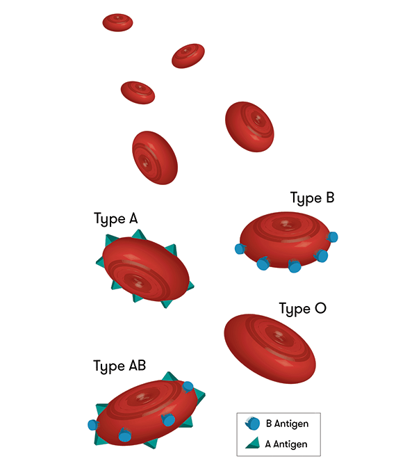
October 3: From Type A to Type O
We all know and love universal blood type O, but what about those who actually have it? For kidney transplants, type-A positive, -B positive, and -AB positive patients can receive their own respective type and type-O; however, type-O patients can only receive type-O kidneys. Thus, when these other patients receive type-O kidneys, people with type-O lack donors, end up waiting two to four years longer for their kidneys, and often die during the wait. Oh, and let’s not forget that type-O patients comprise over half of the kidney waiting lists!
Scientists from the University of British Columbia have been tirelessly studying this catastrophe for over a decade, and they published their first successful transplant this October 3. They managed to place two reactive enzymes in a type-A kidney so that the kidney changed to universal type-O. Sugars that coat organs’ blood vessels determine blood type, so they created an enzyme reaction to strip away the defining sugars. While past conversions have needed live donors and changed antibodies within patients, compromising their immune systems, this new method changes the kidney itself and uses deceased donors.
So, here’s what happened in their transplant test:
Scientists converted a type-A kidney using the enzymes
Placed the kidney in a deceased recipient (with the family’s permission)
Days 1-2: the body showed no signs of rejecting the kidney
Day 3: a few of the type-A attributes reappeared, which is a slight reaction, but nothing as severe as in previous conversions
The body showed signs of tolerating the kidney anyway
Success!
October 6: 2025 Nobel Prize in Physiology or Medicine
This year, the 2025 Nobel Prize in Physiology or Medicine has been awarded to three people! Mary E. Brunkow, Fred Ramsdell, and Shimon Sakaguchi earned it for their advancements on ‘peripheral immune tolerance’, the mechanism that ensures the immune system doesn’t hurt the body. Essentially, peripheral immune tolerance prevents humans from having all kinds of autoimmune diseases. However, prior to these three, scientists had no real understanding of why or how this system worked. Brunkow, Ramsdell, and Sakaguchi built on each other’s findings to discover ‘regulatory T cells’, the agents behind peripheral immune tolerance.
1995: Sakaguchi debunked the popular theory of ‘central tolerance’ by discovering a new group of immune cells.
2001: Brunkow and Ramsdell explained why a certain type of mice was particularly defenseless against autoimmune diseases. They found that strain to have a mutation in what they dubbed their ‘Foxp3’ gene, and they showed that humans have a similar gene, which also causes an autoimmune disease when mutated.
2003: Sakaguchi showed that the Foxp3 gene dictates the growth of the cells he previously found. These cells became known as ‘regulatory T cells’, and they supervise cells in the immune system as well as the immune system’s tolerance of the human body.
All this is awesome, but let’s see how their discovery actually impacted modern medicine. Scientists have found that regulatory T cells can actually protect tumours from the immune system, so, in this case, they are looking for a way to dismantle the cells. However, to combat autoimmune diseases, scientists can implant more regulatory T cells into the body to help prevent the immune system from attacking the body. So, just as Ann Fernholm proclaimed, “they have thus conferred the greatest benefit to humankind.”
October 7: 2025 Nobel Prize in Physics
Get this: another trio received the 2025 Nobel Prize in Physics! The Royal Swedish Academy of Sciences bestowed the honor onto John Clarke, Michel H. Devoret, and John M. Martinis for their experiments demonstrating quantum physics within a larger system. Quantum physics, or quantum mechanics, allows tunneling, which is when particles pass through barriers. Normally, the effects of quantum mechanics become negligible once they start working with large particles, but Clarke, Devoret, and Martinis showed that tunneling can still happen in a larger system.
Just like with our last trio, here’s how they did it:
1984-1985: They experimented with passing a current of charged particles through a controlled circuit containing superconductors. They found that the multiple particles acted like one large particle when going through the superconductor. The quantum part of this was that the system used tunneling to go from zero-voltage to a voltage. So, they concluded that quantum mechanics can still cause tunneling in a macroscopic system.
And why do we care? Well, Olle Eriksson, the Chair of the Nobel Committee for Physics, said, “It is wonderful to be able to celebrate the way that century-old quantum mechanics continually offers new surprises. It is also enormously useful, as quantum mechanics is the foundation of all digital technology.” I don’t know about you, but I think I’ll take his word for it.
October 8: 2025 Nobel Prize in Chemistry
Our LAST Nobel Prize trio of October comes in Chemistry! Susumu Kitagawa, Richard Robson, and Omar M. Yaghi received the 2025 Nobel Prize in Chemistry from the Royal Swedish Academy of Sciences for their ‘metal-organic frameworks (MOFs)’. These frameworks are from their new molecular construction, where carbon-based molecules link together metal ions so that the two form MOFs, which are essentially porous crystals. Scientists can then manipulate these MOFs to take in and guard particular substances. MOFs can also create chemical reactions and direct electricity. So, with these MOFs, scientists can design materials with particular functions of their choosing.
1989: Robson began testing the properties of atoms by combining copper molecules with four-pronged molecules, and this created porous crystals similar to MOFs. However, these MOF impersonators were unstable and needed someone to fix them.
1992-2003: Enter- Kitagawa and Yaghi. From his experiments, Kitagawa concluded that MOFs could be changed and modified as gases could run through them. Then, Yaghi made a stable MOF and showed that they could be manipulated to have new properties.
Since their discoveries, scientists have made tons of their own unique MOFs, each equipped to solve a different problem. We can thank MOFs for giving us a safer Earth. I mean, any kind of chemical substance that can make clean water, grab carbon dioxide from the air, or produce water from desert air sounds like a good one to me.
October 11: The Surprising Link Between COVID-19 and Anxiety
Covid. The word that teleports Gen-Z right back to online school in pajamas, Roblox, and Charli D’Amelio. We all know and hate it, but did we realize that it might be affecting future generations who weren’t even alive in 2020?
A study published on October 11 revealed that male mice who contracted COVID-19 birthed children with more anxiety-like behaviors than those of uninfected mice’s children. Basically, COVID-19 changes RNA molecules in the male’s sperm, which then dictates his children’s brain development. In female offspring specifically, their brain’s hippocampus region, which deals with behaviors including anxiety and depression, was altered. The authors of the study believe that these changes may cause increased anxiety levels.
Okay, okay. Remember: this study was done on mice, not humans. More research is needed to see if humans will experience similar effects, but for now, we’re safe.
October 12: Light Years Away
“A long time ago in a galaxy far, far away…” Wait, what? A long time ago? Evidence suggesting that the closest alien civilization may be 33,000 light-years away did come out this October 12, but for the estimate to be feasible, the civilization would need to have already existed for at least 280,000 years. Yeah, that feels like a long time ago. And don’t worry about the far, far away part– I’d call 33,000 light-years pretty far.
At a recent meeting in Helsinki, research was shown indicating such a possibility. Here’s the criteria for a planet to have extraterrestrial life and actually sustain itself:
Carbon dioxide in the atmosphere (so photosynthesis can work and support life)
An atmosphere of at least 18% oxygen (complex animals need more oxygen, and there must be enough oxygen for fire because blacksmithing must happen to technologically advance)
Average lifetime of about 10 million years (so they can exist at the same time as us)
Already existed for at least 280,000 years (so civilization can develop and they can exist at the same time as us)
Keeping these in mind, scientists have concluded that if there is an alien civilization existing at the same time as us in the same galaxy, it would have to be at least 33,000 light-years away. To put that into perspective, our Sun is about 27,000 light-years away from us. Yeah. Pretty far.
October 20: Enteral Ventilation
Sometimes, CPR isn’t enough to save respiratory failure. Then, patients turn to mechanical ventilation. But sometimes mechanical ventilation is too much, and the lungs end up even further damaged. Enteral ventilation, however, may just be the sweet spot. Enteral ventilation is a practice where perfluorodecalin, an exceptionally oxygen-soluble liquid, is administered through the intestine to deliver oxygen to the body while the lungs heal. Published on October 20, the first in-human study of enteral ventilation succeeded and was demonstrated to be safe. The only side effects were bloating and stomach pain, but those quickly resolved, and perfluorodecalin concentrations nearly disappeared from the bloodstream (a good thing!).
After this safe and tolerated success, more studies on enteral ventilation will soon develop, and lungs everywhere may be saved.
October 20: CI Chondrite on the Moon
Before we get into any of this moon stuff, you may be wondering what in the world (or should I say galaxy) CI Chondrite is. I’m here to help! CI Chondrite, a porous and the most water-dense meteorite, generally breaks before it can reach Earth because its properties make it so crumbly. CI Chondrite actually makes up less than one percent of all meteorites on Earth. That means it also barely ever reaches the moon. However, during their Chang’e-6 mission published on October 21, the China National Space Administration found traces of CI Chondrite dust on the moon.
A Chondrite Meteorite
Here’s how they did it:
They looked at thousands of fragments from the Apollo Basin, a sub-basin in the South Pole-Aikten Basin that acts as a hotspot for debris since it covers one-fourth of the moon.
They looked for pieces with olivine, a mineral normally in meteorites.
Then, they analyzed the olivine pieces and found seven with properties identical to CI Chondrite
When analyzing, they found that the pieces did not have the chemical ratios expected for lunar debris.
However, they realized that the seven fragments’ ratios did align with those of a CI Chondrite asteroid that crashed, melted, and solidified on the moon early in the solar system’s history.
With these discoveries, the team found the first solid evidence that CI Chondrite once hit the moon and that CI Chondrite can be preserved after such a crash. Actually, they found that CI Chondrite could comprise up to 30 percent of the Moon’s meteorite debris. Additionally, their study provided evidence to help back up the theory that CI Chondrite once created water and volatiles on the Earth and Moon. More research is needed to see if it’s really true, but those missions will now be much easier with the China National Space Administration’s new process to find CI Chondrite.
October 27: Back to the Basics
Nope, not like the song. On October 27, in the Astrophysical Journal Letters, scientists described their findings of what they believed to be Population III stars, one of the first groups of stars in the galaxy. With the James Webb Space Telescope, they pinpointed them in LAP1-B, a cluster of stars 12 billion light-years away from Earth. Scientists believe Population III stars are some of the first stars made after the Big Bang, and they have a unique property of being a billion times brighter than and a million times the mass of our Sun.
Here’s why they believe their discovered stars to be Population III:
Emission lines on the stars’ spectra indicated high-energy photons, which are consistent with Population III stars.
Their spectra showed them to be extremely large.
Their masses aligned with astronomers’ guesses for those of Population III stars.
They were in LAP1-B, whose properties agree with the criteria for Population III.
It’s a low hydrogen and helium environment.
Its temperature can support star formation.
It’s a low-mass cluster, and it had few large stars before those of Population III.
It meets mathematical criteria for forming stars and keeping them alive.
Seems pretty feasible, right? Anyways, these scientists were the first to find a group of stars that meets all criteria for being Population III, and these ancient stars can actually explain the galaxy’s construction and development. That’s all for STEM this October, but don’t worry, because this November’s looking like a great one.
University of British Columbia. (2025, October 3). UBC enzyme technology clears first human test toward universal donor organs for transplantation. Eurek Alert. https://www.eurekalert.org/news-releases/1100223
How Scientists are Using Worms to Learn About Humans
Worms and humans could not possibly be any more different. And yet, scientists have been studying C. elegans (caenorhabditis elegans) to learn more about the human body over 70 years. These unassuming worms have aided in groundbreaking findings in medicine for human diseases such as Alzheimer’s, AIDS, and stroke.
What makes C. elegans so valuable is not its complexity, but rather its simplicity. Because so many of its biological pathways are conserved in humans, this worm provides a uniquewindow into the fundamental processes of life, including cell division, gene regulation, neural signaling, and aging. With a transparent body, rapid life cycle, and a genetic makeup that mirrors much of our own, C. elegans has become an essential organism in modern biomedical research. Understanding how scientists use these worms can help us appreciate not just what we’ve already learned, but also the vast potential that still lies ahead.
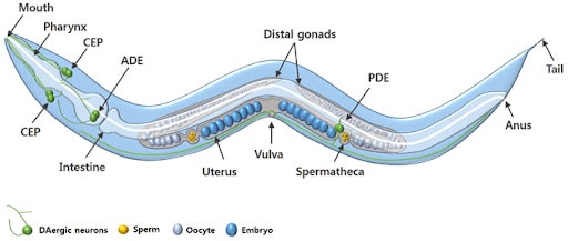
What is C. elegans?
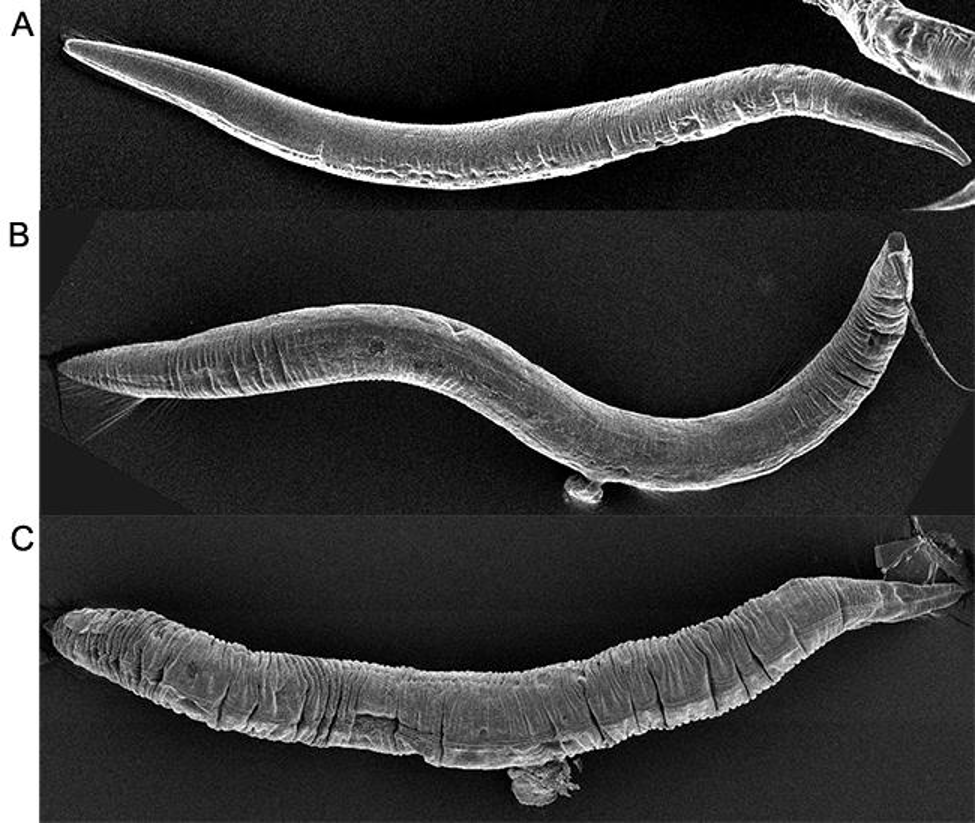
C. elegans is a free-living nematode that has become one of the most important model organisms in research. It measures approximately one millimeter in length and naturally lives in temperate soil environments, where it feeds on bacteria like e. coli. It is non-parasitic and exists in two sexes: hermaphrodites, which are capable of self-reproduction, and males, which occur at a less than 0.1% chance under normal conditions. The hermaphroditic reproductive mode allows for the maintenance of isogenic populations, which is advantageous for genetic studies.
The adult C. elegans hermaphrodite has exactly 959 somatic cells while the adult male C. elegans has exactly 1,031 somatic cells. The worm’s relatively simple anatomy includes muscles, a nervous system, a digestive system, a reproductive system, and an excretory system. The organism develops through four larval stages before reaching adulthood, with a complete lifecycle taking just two to three weeks under laboratory conditions.
Genetically, C. elegans has a compact genome consisting of about 100 million base pairs across six chromosomes. It was the first multicellular organism to have its entire genome sequenced in 1998 in a project led by John Sulston and Bob Waterstons. Its genome is highly amenable to manipulation using a variety of modern techniques.
Why do scientists study C. elegans specifically?
First introduced into studies by Sydney Brenner in the 1960s to study neurological development and the nervous system, the nematode proved itself in the lab with its unique combination of genetic, anatomical, and practical features that make it exceptionally suitable for biomedical research.
Remarkably, around 60-70% of human disease-associated genes have counterparts in the C. elegans genome, making it an incredibly valuable model for studying human biology. Many genes responsible for critical cellular functions are evolutionarily conserved between worms and humans. Therefore, scientists can manipulate the function of these genes in C. elegans to study their roles in disease without the complexity or ethical challenges of working with human subjects or higher animals like mice or primates.
Adult hermaphrodites’ cells, which remain the same in every single worm, each of which has been identified and mapped, allowing for detailed tracking of development, differentiation, and cellular processes. Its transparent body enables real-time visualization of internal structures, including neurons, muscles, reproductive organs, and digestive tissues. The worm, which has a simple nervous system of only 302 cells, is one of the only organisms where every neural connection is known. Additionally, C. elegans has a short life cycle of two to three weeks and is easy to culture in large numbers, making it especially convenient for developmental and aging studies.
How do scientists modify C. elegans in experiments?
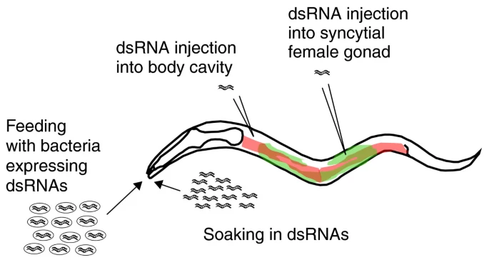
Scientists modify and study C. elegans using four primary methods: RNA interference (RNAi), CRISPR-Cas9 genome editing, transgenic techniques, and drug screening.
One of the most widely used techniques for modifying gene expression in C. elegans is RNA interference (RNAi). This method allows scientists to silence specific genes to observe the effects of their absence. In C. elegans RNAi can be easily administered by feeding worms with genetically engineered E. coli bacteria that produce double-stranded RNA (dsRNA) matching the gene of interest. Once ingested, the dsRNA activates the worm’s endogenous RNAi pathway, leading to the degradation of the corresponding messaging RNA and a reduction or elimination of the target protein. This method is highly efficient, non-invasive, and relatively easy to perform, making it ideal for large-scale genetic screens. Researchers can identify genes involved in key processes such as embryonic development, aging, metabolism, and neurodegeneration.
The CRISPR-Cas9 system has revolutionized genetic research in C. elegans by enabling precise, targeted alterations to the genome. Scientists introduce a complex composed of the Cas9 enzyme and a guide RNA (gRNA) into the worm, which directs the Cas9 to a specific DNA sequence. Once there, Cas9 introduces a double-strand break in the DNA. The cell’s natural repair mechanisms then fix the break, and researchers can insert, delete, or replace specific DNA sequences. In C. elegans, CRISPR can create mutants mimicking human disease alleles or study regulatory elements of genes. This method provides a level of control that surpasses RNAi, as it allows for permanent and heritable genetic modifications. Scientists often inject the CRISPR-Cas9 components directly into the gonads of adult hermaphrodites, ensuring that the genetic changes are passed onto the offspring.
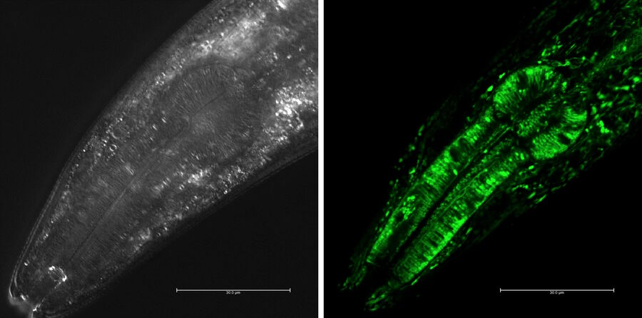
Transgenic techniques in C. elegans insert foreign DNA into the worm’s genome to monitor gene expression, trace cell lineages, or study protein localization. One common approach is to fuse a gene of interest to a reporter gene such as green fluorescent protein (GFP). When this gene is expressed, the fluorescent tag can be visualized in living worms using fluorescence microscopy. This allows researchers to observe where and when specific genes are active, how proteins move within the cells, and how cells interact during development or disease progression. Transgenes are typically introduced via microinjection into the syncytial gonads of adult worms, leading to the formation of extrachromosomal arrays inherited by the next generation. Stable lines can also be created through CRISPR or chemical integration methods. These visual tools are particularly powerful due to the worm’s transparent body, which makes it possible to track fluorescent signals in real time throughout the entire organism.
C. elegans is an excellent system for drug screening and environmental toxicology due to its small size, short lifespan, and genetic tractability. Researchers can test the effects of thousands of compounds quickly and cost-effectively. In these experiments, worms are exposed to chemical agents in liquid or on agar plates, and their survival, movement, reproduction, or specific cellular markers are measured to assess the biological impact. Using genetically modified strains that mimic human disease pathways, scientists can screen for drugs that alleviate symptoms or restore normal function. These tests provide an efficient first step in drug development, singling out promising candidates before moving onto mammalian models.
The cell lineage and the programmed cell death in C. elegans / Nobel Prize in Physiology or Medicine 2002
One of the most groundbreaking discoveries made using C. elegans was the genetic basis of programmed cell death, or apoptosis, a critical process in both development and disease. The research was led by Dr. H. Robert Horvitz at the Massachusetts Institute of Technology. Horvitz and his colleagues began studying cell death in C. elegans in the 1980s by tracing the fate of every cell in the worm’s body during development. They discovered that exactly 131 cells always die in the developing hermaphrodite and that this process was genetically controlled. Through genetic screening, Horvitz identified three core genes that regulated apoptosis: ced-3, ced-4, and ced-9. By inducing mutations in these genes, the researchers could either prevent or accelerate cell death in the worm. This revealed that cell death is not a passive consequence of damage, but rather an active, genetically programmed event. The mammalian counterparts of these genes, like caspases and BCL-2, were later discovered to play central roles in cancer, autoimmune diseases, and neurodegeneration, making this research foundational to modern medicine. Horvitz was awarded the 2002 Nobel Prize in Physiology or Medicine for his work along with Sydney Brenner and John Sulston.
In addition, C. elegans has contributed to our understanding of neurodegenerative diseases such as Alzheimer’s. One major study was led by Dr. Christopher Link at the University of Colorado in the late 1990s. Link developed a transgenic C. elegans strain that expressed the human β-amyloid (Aβ) peptide in muscle cells. This is the same peptide that forms toxic plaques in the brains of Alzheimer’s patients. In the study, the researchers observed that worms expressing Aβ developed progressive paralysis as they aged, mimicking aspects of human Alzheimer’s pathology. They then used this model to screen for genetic mutations and chemical compounds that could suppress the toxic effects of Aβ. Their work identified several genes involved in protein folding and stress response that modified Aβ toxicity. This demonstrated that C. elegans could be used as a fast and cost-effective in vivo system for identifying genetic and pharmacological modifiers of Alzheimer’s disease. The worm model has since then been adapted by numerous labs worldwide to study tau protein aggregation and mitochondrial dysfunction, expanding our knowledge of neurodegenerative pathways.
Another major discovery made using C. elegans was the link between insulin signaling and lifespan regulation. Dr. Cynthia Kenyon at the University of California, San Francisco, led a series of experiments in the 1990s that transformed the field of aging research. Kenyon’s team discovered that a single mutation in the daf-2 gene, which encodes an insulin/IGF-1 receptor, could double the worm’s lifespan. They found that when daf-2 signaling was reduced, it activated another gene, daf-16, which promoted the expression of stress-resistance and longevity-related genes. To test this, Kenyon used genetic mutants and tracked their development and survival across generations. The C. elegans with the daf-2 mutation lived significantly longer than their wild-type counterparts and were more resistant to oxidative stress and heat. These findings provided the first clear evidence that aging could be actively regulated by specific genetic pathways rather than being a passive deterioration process. Later studies found that similar insulin/IGF-1 pathways exist in mammals, including humans, opening new therapeutic avenues for age-related diseases, diabetes, and metabolic disorders.
So what does the future hold?
The future of C. elegans in scientific research is remarkably promising, with its applications continually expanding as technology and genetic tools advance. With the rise of CRISPR-Cas9, optogenetics, and high-throughout screening techniques, researchers can now manipulate C. elegans with unprecedented precision to study complex biological processes such as epigenetics, gut-brain interactions, and real-time neuronal activity. In the coming years, C. elegans is expected to play an even greater role in personalized medicine and systems biology. Its potential as a predictive model for human gene function could aid in understanding the consequences of mutations found in patient genomes, leading to more tailored treatments. The worm’s short life cycle, fully mapped genome, and conserved biological pathways make it an ideal model for rapidly identifying new therapeutic targets and testing drugs, especially for age-related and neurodegenerative diseases. Despite its simplicity, this tiny nematode continues to open doors to complex human biology, proving that even the smallest organisms can have the biggest impact on science and medicine.
“C. Elegans 101: A White Paper – InVivo Biosystems.” InVivo Biosystems, 26 Jan. 2024, invivobiosystems.com/disease-modeling/c-elegans-101-a-white-paper/.
Edgley, Mark. “What Is Caenorhabditis Elegans and Why Work on It? – Caenorhabditis Genetics Center (CGC) – College of Biological Sciences.” Umn.edu, University of Minnesota, 2022, cgc.umn.edu/what-is-c-elegans.
Venkatesan, Arun, and Krishma Adatia. “Anti-NMDA-Receptor Encephalitis: From Bench to Clinic.” ACS Chemical Neuroscience, vol. 8, no. 12, 7 Nov. 2017, pp. 2586–2595, https://doi.org/10.1021/acschemneuro.7b00319.
Wheelan, Sarah J, et al. “Human and Nematode Orthologs — Lessons from the Analysis of 1800 Human Genes and the Proteome of Caenorhabditis Elegans.” Gene, vol. 238, no. 1, Sept. 1999, pp. 163–170, https://doi.org/10.1016/s0378-1119(99)00298-x.
“Whitehead Institute of MIT.” Whitehead Institute of MIT, wi.mit.edu/unusual-labmates-how-c-elegans-wormed-its-way-science-stardom.
“Wonderous Worms.” NIH News in Health, 3 July 2025, newsinhealth.nih.gov/2025/07/wonderous-worms. Accessed 1 Aug. 2025.
Zhang, Siwen, et al. “Caenorhabditis Elegans as a Useful Model for Studying Aging Mutations.” Frontiers in Endocrinology, vol. 11, 5 Oct. 2020, https://doi.org/10.3389/fendo.2020.554994.
Huntington’s Disease, discovered by George Huntington in 1872, is a hereditary genetic brain disorder. Since then, many researchers have dedicated their lives to studying Huntington’s Disease. While we have not found a cure nor treatments to slow the progression, we have discovered how it works, what it is, what it can do, and how it is passed down.
George Huntington, an American physician from Long Island with a degree from Columbia University, published his paper “On Chorea” in 1872, describing Huntington’s Disease so accurately and succinctly that the disease was named after him. He was only 21 when his paper was published. However, he first encountered what would come to be known as Huntington’s Disease when he was 8 years old while accompanying his father and grandfather on medical rounds. Within “On Chorea”, he summarized three key characteristics of a person with Huntington’s Disease: their propensity to suicide and mental disorders, inheritance patterns, and progressive disabilities. This was his sole contribution to medical research. His paper shone a light on this “medical curiosity” from a new field of medicine and shook the medical research world into a frenzy to try to grasp what Huntington’s was and how it worked.
Huntington’s Disease (HD), is inherited from your parents following an autosomal dominant inheritance pattern. It causes nerve cells, mainly in the basal ganglia, brain cortex, and the striatum, to gradually break down and lose function. More than 15,000 Americans currently have HD, but many more are at risk of developing it. There are two kinds of Huntington’s Disease, adult onset, the most common, and early onset, which affects children and teenagers. Fortunately, early onset is very rare, only affecting 5.7% of Huntington’s cases. HD affects an estimated 3 to 7 people out of 100,000, most commonly people of European descent. If a parent has HD, their child has a 50% chance of inheriting the genetic mutation as well. If the child does not inherit it, they will not show symptoms and cannot pass it down. On the condition that the patient has more than 50 CAG repeats, there is a 90% chance they inherited the gene from their father, because CAG repeats tend to be more unstable when passed from the male. There are situations where HD occurs without family history. This event is called Sporadic HD.
Huntington’s is a genetic mutation of the HTT gene. It produces a protein called huntingtin. This protein helps your nerves function. The HTT gene is found on chromosome 4, which also happens to be associated with the cause of many other genetic disorders and some types of cancer. The defect involves a DNA segment known as CAG trinucleotide repeat. It is made up of three DNA building blocks, cytosine, adenine, and guanine, appearing several times in a row. Normally, the CAG segments are repeated 10 to 35 times within a gene, and these people lie in the unaffected range, whether normal or intermediate allele sub-ranges. To a person with Huntington’s, it can be repeated 36 to more than 120 times. They lie in the affected range, either reduced penetrance or full penetrance if they have more than 40 CAG repeats. People in the intermediate allele and the reduced penetrance sub-ranges, with 27-39 CAG repeats, may not develop symptoms but can be carriers. The increase in repeats leads to the production of abnormally long and oddly shaped huntingtin proteins. The elongated protein forms toxic fragments that fuse together and collect in neurons, disrupting the normal function of cells and ultimately killing them. This causes the symptoms of Huntington’s Disease. As the mutated HTT gene is passed down, the amount of CAG trinucleotide repeats increases. A larger number of repeats causes early onset Huntington’s and a sooner appearance of symptoms. This is referred to as anticipation.
The diverse symptoms of Huntington’s Disease are what leads to many misdiagnoses in the early stages and why it took so long to be recognized as its own disease. George Huntington’s paper “On Chorea” focused mostly on chorea, which involves involuntary jerking or writhing movements, akinesiadeveloping as the disease progresses, unusual or slow eye movements, trouble with walking and balance, dystonia, ataxia, trouble with speech, athetosis, and dysphagia, and weight loss. Mental health conditions include irritability, mood swings, social withdrawal, insomnia, fatigue, loss of energy, suicidal thoughts, OCD, mania, bipolar disorder, psychosis, hallucinations, and paranoia. There are cognitive conditions as well, like, trouble organizing, trouble prioritizing and focusing on tasks, lack of flexibility and perseveration, lack of impulse control that can lead to violent outbursts, lack of awareness in one selves behaviors and ability, slowness in processing thoughts, seizures, trouble with driving, and trouble learning new information and memorization. These symptoms can get more intense when the person is nervous or distracted. Eventually, these symptoms get so bad that it is more closely categorized as dementia.
Many people with HD remain conscious of their environment and can express emotions. As it progresses, the patient will need more help and supervision. Ultimately, they will need help at all hours of the day. HD is not fatal in and of itself. Patients most commonly die from complications like physical injury from falls and accidents, malnutrition due to trouble feeding oneself, infections, typically pneumonia but others as well, choking, heart failure, seizures, and, due to the mental toll, 7-10% of HD patients commit suicide. The average lifespan of a person with Huntington’s is 10 to 30 years after a diagnosis.
This disease, because of its diverse symptoms, takes a skilled eye to diagnose. In most cases, it can be done with a neurological exam and an analysis of the patient’s medical and family history. But in other cases, the patient might require genetic and blood tests and diagnostic imaging like an MRI, CT, PET scan, or EEG. A neurologist and neuropsychiatrist will perform these tests. There are many research studies underway to study Huntington’s and while we do not have a cure, we have a basic understanding of the disease, which means we are one step closer to long term treatments. Johns Hopkins, for example, has 4 ongoing studies: the Sage Studies: PERSPECTIVE Program, which is evaluating the safety and efficiency of the experimental drug SAGE-718 in adults with early Huntington’s Disease, the Generation HD2 tests, which is the second phase of tests on Tominersen in young adults with HD ranging from 25-50 years old. The HDClarity study, an observational study to collect cerebrospinal fluid in order to study biomarkers that influence HD’s pathophysiology and growth, and the Enroll-HD program, a registry for the Global Huntington Disease Cohort, providing vast information for future clinical research. These are just a few of the many programs dedicated to unlocking the mysteries of HD. The most promising fields are those studying biomarkers, like the HDClarity study, and stem cell research.
There are many options for treatments that can help improve the quality of life for a person with HD. They will require more help as the disease progresses and a team of people to help them like a neurologist, psychiatrist, genetic counselor, physical therapist, occupational therapist, and a speech therapist. A counselor could also help the patient and their family members with the emotional toll. Medications can also be prescribed to ease symptoms and keep them functioning as long as possible. To treat chorea they could take deutetrabenazine, amantadine, tetrabenazine, or haloperidol. The latter two of which could also help deter hallucinations and delusions. To manage their emotions, they could be prescribed antidepressants like fluoxetine and sertraline, antipsychotic drugs like risperidone and olanzapine; however, some antipsychotic medications have side effects that could make chorea and akinesia worse, and mood stabilizing medications like lithium. Antidepressant and antianxiety medications are also commonly prescribed because there are high rates of depression and suicide amongst patients with HD. It is also recommended to maintain physical fitness because it is shown that patients who exercise regularly delay the symptoms of HD more than those who do not. Huntington’s, however, can be prevented by genetic counseling, prenatal testing, and in vitro fertilization, where an egg and sperm are fertilized in a lab and checked to see if it has Huntington’s disease. If it does not, it is then implanted back into the uterus. It is important to speak to a genetic counselor before having a child if you or your partner has HD or is at risk to develop symptoms.
An HD diagnosis is certainly not a death sentence. A person with Huntington’s can live a long, happy life. We now know so much about this disease that even George Huntington would not be able to believe. There are many options for every particular patient and every particular case. And as science and technology advances, so will we in our path to finding a cure for Huntington’s Disease.
Glossary
1. A CAG trinucleotide repeat is an unstable expansion of the DNA sequence “cytosine-adenine-guanine” (CAG) that codes for the amino acid glutamine, resulting in a long “polyglutamine” tract within a protein
2. a situation where individuals who inherit a disease-causing genetic mutation do not develop the associated disease or condition
3. Akinesia: become rigid (stiff) and move very little or not at all
4. Dystonia: unusual fixed (unchanging) postures
5. Ataxia: loss of coordination
6. Athetosis: slow, involuntary, and writhing movements
7. Dysphagia: difficulty swallowing
8. Psychosis: losing some contact with reality
9. Tominersen: a treatment for Huntington’s Disease that is under research and trials
Earlier this summer, I was graciously given the opportunity to shadow a private-practice oncologist/hematologist in the Dallas area. There, I gained a clear understanding of what a career in STEM entails, learned how doctors approach complex cancer cases, and secured an inside view into the emotionally taxing yet deeply rewarding work of an oncologist.
What does an Oncologist’s career look like?
At the ground level, an oncologist’s job involves diagnosing and treating cancer. They play a central role in administering cancer treatments and developing long-term plans. There are three main types of oncologists:
Medical Oncologist: Dr. Nair, whom I shadowed, practices as a medical oncologist. These doctors use targeted therapies like chemotherapy and immunotherapy to treat cancers.
Surgical Oncologist: Surgical oncologists perform biopsies and remove tumors through surgical procedures. Usually, after a medical oncologist has successfully shrunk a tumor through targeted therapy, a surgical oncologist will excavate the remaining piece.
Radiation Oncologist: As the name suggests, these doctors treat cancer through radiation therapy.
Dr. Nair works as a hematologist-oncologist. Because cancer often involves blood and bone marrow (leukemia, lymphoma, myeloma), having training in both oncology (solid tumors) and hematology (blood disorders) allows a doctor to treat a wider variety of patients without having to refer them to another clinic. Also, in the U.S., most oncologists need no extra schooling to end up board-certified in both.
Typically, becoming an oncologist requires about 14-16 years of school. This includes a four-year undergraduate program, where students generally major in biology, chemistry, mathematics, or physics. Then, students take the MCAT, or the Medical College Admission Test, and attend medical school to earn their MD. After four years of medical school, doctors attend a three-year residency program. Finally, they complete a three-year fellowship program, subspecializing in oncology or hematology-oncology. Oncologists typically finish schooling in their mid-thirties, and though they spend most of their twenties in schooling, many agree that this time is fully necessary due to the extensive information students have to understand.
A central part of an oncologist’s job is responding to a wide spectrum of questions, ranging from emotional ones like “if the tumor is getting bigger, do I have less time to live?” to straightforward questions like, “if I eat and sleep more, will I have more energy the next morning?” Sure, many of these questions become routine over time, but it’s that rare, complex one that truly tests a doctor’s knowledge and, when answered well, builds even more trust between the patient and their provider. Because cancer is such a serious topic, patients seek oncologists who make them comfortable, and the best way to provide that security is by easing their uncertainties and reinforcing confidence in their provider. This is exactly why those 14 long years of medical training matter so much.
The Difference Between Private Practice and Clinic
Dr. Nair is affiliated with the broader group Texas Oncology and practices at Medical City Dallas, but before going in to shadow her, I had no idea what the difference between a private practice and a clinic was. Here is an easy way to break it down:
Private practice: When a doctor or group of doctors owns, manages, and runs their own medical office. Like a business, they hire staff, manage billing, and run their own practice from top to bottom. Though private practice intersects the two contrasting fields of medicine and business, these doctors have more flexibility when not working for a large hospital or healthcare system.
Clinic: Usually affiliated with a larger group, hospital, or university. Doctors who work as part of a clinic follow the protocol set up by a broader employer and focus less on business and management.
Highlight Patients
You may think that looking at cancer gets repetitive after a while, and maybe you’re right- but in the two weeks that I shadowed Dr. Nair, we saw a wide variety of patients that kept me quite interested. Often, it wasn’t the cancer or condition that made them memorable, but their personality, and the reminder that cancer does not discriminate. People from all walks of life, rich or poor, tall or short, male or female, can be struck by the disease at random and affected in similar ways.
1. Female, mid-40s, obese
This patient was on blood-thinners that were administered by the hospital. Upon arriving home, she purposefully took double the prescribed dose for a few days. With the alarmingly high dosage this patient was taking, her gums would bleed when brushing her teeth, and minor cuts would bleed profusely without stopping. Suddenly, the patient formed a massive internal hemorrhage in her stomach, and was rushed to the ICU where she took a break from blood thinners and recuperated.
2. Female, mid-30s
This patient was aware she had a tumor in her lungs, but didn’t know the extent of its spread or whether it was even malignant. As the cardiothoracic surgeon opened her chest to perform a biopsy and assess the situation, he found that the cancer presented as stage 4 and had spread extensively throughout the lungs. After removing substantial diseased lung tissue, the patient’s remaining lung capacity was too low to sustain oxygenation. Therefore, she was placed on a ventilator that essentially acted as a pair of bedside lungs, pumping air for her.
3. Female, early-60s, groaning in pain
As Dr. Nair and I walked into the patient’s room, she was lying on the bed, groaning and screaming in severe pain. This woman had a pancreatic tumor, one of the most painful types of cancer, due to the tumor pressing on bunches of nerves and organs in the abdomen and back. Though she was fully lucid, the pain was preventing her from formulating complete thoughts or ideas, and her husband described that she could not eat properly or move around without a wheelchair. Dr. Nair told the couple to visit the ER within the hospital immediately, so that the patient could be administered stronger pain medications.
The role of women in healthcare
One thing that really stuck out to me was the number of women who worked in the office with Dr.Nair. Out of the three oncologists, only one was a man, and the rest of the staff, including the P.A. and infusion nurses, were all women.
In fact, according to the U.S. Bureau of Labor Statistics; around 77.6% of all healthcare workers are women. However, we hold a disproportionately small number of leadership positions compared to men. Where 77.6% of healthcare workers are women, only about 38% of all physicians are women.
Despite the gender gap that still exists today, equality growth in the last 20 years alone has been monumental. According to the Association of American Medical Colleges,
“From 2004 to 2022, the number of women in the active physician workforce increased 97%.”
Going forward, the future looks bright too. In 2019, women for the first time accounted for a majority (50.5%) of students enrolled in medical school in the United States. Today, women account for about 54.6% of medical school students. As women make up the majority of medical school graduates, the number of physicians in the coming years will consequently increase.
Conclusion
Before I arrived at the oncologist’s office, I pictured a gloomy waiting room filled with silent, dejected patients. Instead, I discovered something completely different. People tend to imagine only the sickest patients at a cancer clinic, the ones who are dying. But they often forget about the many who are improving, on the uphill climb, and who see the doctor’s office not as a place of punishment or despair, but as a lifeline that offers hope and light at the end of the tunnel.
Seeing this side of cancer care reshaped my view of healthcare entirely. It made me realize that medicine isn’t just about treating disease and sending patients on their way, but instead creating an environment where people are given a reason to keep fighting.