
Earlier this summer, I was graciously given the opportunity to shadow a private-practice oncologist/hematologist in the Dallas area. There, I gained a clear understanding of what a career in STEM entails, learned how doctors approach complex cancer cases, and secured an inside view into the emotionally taxing yet deeply rewarding work of an oncologist.
What does an Oncologist’s career look like?
At the ground level, an oncologist’s job involves diagnosing and treating cancer. They play a central role in administering cancer treatments and developing long-term plans. There are three main types of oncologists:
Medical Oncologist: Dr. Nair, whom I shadowed, practices as a medical oncologist. These doctors use targeted therapies like chemotherapy and immunotherapy to treat cancers.
Surgical Oncologist: Surgical oncologists perform biopsies and remove tumors through surgical procedures. Usually, after a medical oncologist has successfully shrunk a tumor through targeted therapy, a surgical oncologist will excavate the remaining piece.
Radiation Oncologist: As the name suggests, these doctors treat cancer through radiation therapy.
Dr. Nair works as a hematologist-oncologist. Because cancer often involves blood and bone marrow (leukemia, lymphoma, myeloma), having training in both oncology (solid tumors) and hematology (blood disorders) allows a doctor to treat a wider variety of patients without having to refer them to another clinic. Also, in the U.S., most oncologists need no extra schooling to end up board-certified in both.
Typically, becoming an oncologist requires about 14-16 years of school. This includes a four-year undergraduate program, where students generally major in biology, chemistry, mathematics, or physics. Then, students take the MCAT, or the Medical College Admission Test, and attend medical school to earn their MD. After four years of medical school, doctors attend a three-year residency program. Finally, they complete a three-year fellowship program, subspecializing in oncology or hematology-oncology. Oncologists typically finish schooling in their mid-thirties, and though they spend most of their twenties in schooling, many agree that this time is fully necessary due to the extensive information students have to understand.
A central part of an oncologist’s job is responding to a wide spectrum of questions, ranging from emotional ones like “if the tumor is getting bigger, do I have less time to live?” to straightforward questions like, “if I eat and sleep more, will I have more energy the next morning?” Sure, many of these questions become routine over time, but it’s that rare, complex one that truly tests a doctor’s knowledge and, when answered well, builds even more trust between the patient and their provider. Because cancer is such a serious topic, patients seek oncologists who make them comfortable, and the best way to provide that security is by easing their uncertainties and reinforcing confidence in their provider. This is exactly why those 14 long years of medical training matter so much.
The Difference Between Private Practice and Clinic
Dr. Nair is affiliated with the broader group Texas Oncology and practices at Medical City Dallas, but before going in to shadow her, I had no idea what the difference between a private practice and a clinic was. Here is an easy way to break it down:
Private practice: When a doctor or group of doctors owns, manages, and runs their own medical office. Like a business, they hire staff, manage billing, and run their own practice from top to bottom. Though private practice intersects the two contrasting fields of medicine and business, these doctors have more flexibility when not working for a large hospital or healthcare system.
Clinic: Usually affiliated with a larger group, hospital, or university. Doctors who work as part of a clinic follow the protocol set up by a broader employer and focus less on business and management.
Highlight Patients
You may think that looking at cancer gets repetitive after a while, and maybe you’re right- but in the two weeks that I shadowed Dr. Nair, we saw a wide variety of patients that kept me quite interested. Often, it wasn’t the cancer or condition that made them memorable, but their personality, and the reminder that cancer does not discriminate. People from all walks of life, rich or poor, tall or short, male or female, can be struck by the disease at random and affected in similar ways.
1. Female, mid-40s, obese
This patient was on blood-thinners that were administered by the hospital. Upon arriving home, she purposefully took double the prescribed dose for a few days. With the alarmingly high dosage this patient was taking, her gums would bleed when brushing her teeth, and minor cuts would bleed profusely without stopping. Suddenly, the patient formed a massive internal hemorrhage in her stomach, and was rushed to the ICU where she took a break from blood thinners and recuperated.
2. Female, mid-30s
This patient was aware she had a tumor in her lungs, but didn’t know the extent of its spread or whether it was even malignant. As the cardiothoracic surgeon opened her chest to perform a biopsy and assess the situation, he found that the cancer presented as stage 4 and had spread extensively throughout the lungs. After removing substantial diseased lung tissue, the patient’s remaining lung capacity was too low to sustain oxygenation. Therefore, she was placed on a ventilator that essentially acted as a pair of bedside lungs, pumping air for her.
3. Female, early-60s, groaning in pain
As Dr. Nair and I walked into the patient’s room, she was lying on the bed, groaning and screaming in severe pain. This woman had a pancreatic tumor, one of the most painful types of cancer, due to the tumor pressing on bunches of nerves and organs in the abdomen and back. Though she was fully lucid, the pain was preventing her from formulating complete thoughts or ideas, and her husband described that she could not eat properly or move around without a wheelchair. Dr. Nair told the couple to visit the ER within the hospital immediately, so that the patient could be administered stronger pain medications.
The role of women in healthcare
One thing that really stuck out to me was the number of women who worked in the office with Dr.Nair. Out of the three oncologists, only one was a man, and the rest of the staff, including the P.A. and infusion nurses, were all women.
In fact, according to the U.S. Bureau of Labor Statistics; around 77.6% of all healthcare workers are women. However, we hold a disproportionately small number of leadership positions compared to men. Where 77.6% of healthcare workers are women, only about 38% of all physicians are women.
Despite the gender gap that still exists today, equality growth in the last 20 years alone has been monumental. According to the Association of American Medical Colleges,
“From 2004 to 2022, the number of women in the active physician workforce increased 97%.”
Going forward, the future looks bright too. In 2019, women for the first time accounted for a majority (50.5%) of students enrolled in medical school in the United States. Today, women account for about 54.6% of medical school students. As women make up the majority of medical school graduates, the number of physicians in the coming years will consequently increase.
Conclusion
Before I arrived at the oncologist’s office, I pictured a gloomy waiting room filled with silent, dejected patients. Instead, I discovered something completely different. People tend to imagine only the sickest patients at a cancer clinic, the ones who are dying. But they often forget about the many who are improving, on the uphill climb, and who see the doctor’s office not as a place of punishment or despair, but as a lifeline that offers hope and light at the end of the tunnel.
Seeing this side of cancer care reshaped my view of healthcare entirely. It made me realize that medicine isn’t just about treating disease and sending patients on their way, but instead creating an environment where people are given a reason to keep fighting.
Advancements in genetic engineering have brought revolutionary tools to the forefront of biotechnology, with CRISPR leading as one of the most precise and cost-effective methods of gene editing. CRISPR, which stands for Clustered Regularly Interspaced Short Palindromic Repeats, allows scientists to alter DNA sequences by targeting specific sections of the genome. Originally discovered as part of a bacterial immune system, CRISPR systems have now been adapted to serve as programmable gene-editing platforms. This paper explores how CRISPR works, its current uses, its future potential, and the ethical considerations surrounding its application in both human and non-human systems.
How CRISPR System Works
The CRISPR-Cas system operates by combining a specially designed RNA molecule with a CRISPR-associated protein, such as Cas9 or Cas12a. The RNA guides the protein to a specific sequence in the genome, where the protein then cuts the DNA. Once the strand is cut, natural repair mechanisms within the cell are activated. Researchers can either allow the cell to disable the gene or insert a new gene into the gap. As described by researchers at Stanford University,
“The system is remarkably versatile, allowing scientists to silence genes, replace defective segments, or even insert entirely new sequences.” (CRISPR Gene Editing and Beyond)
This mechanism has been compared to a pair of molecular scissors that can cut with precision. For example, the Cas9 protein is programmed with a guide RNA to recognize a DNA sequence of about 20 nucleotides. Once it finds the target, it makes a double-stranded cut. The repair process that follows enables gene knockouts, insertions, or corrections. This technology has dramatically reduced the time and cost associated with gene editing, making previously complex tasks achievable in weeks rather than months. According to a 2020 review,
“CRISPR/Cas9 offers researchers a user-friendly, relatively inexpensive, and highly efficient method for editing the genome.” (Computational Tools and Resources Supporting CRISPR-Cas Experiments)
CRISPR’s influence extends across many fields, but its role in medicine has attracted the most attention. Scientists are using CRISPR to treat genetic diseases such as sickle cell anemia by editing patients’ own stem cells outside the body and then reinserting them. In 2023, researchers published results showing that a single treatment could permanently alleviate symptoms for some patients with these genetic diseases (Zhang 4.) Another area of exploration includes its potential for treating cancers by modifying immune cells to better recognize and destroy cancerous tissue. According to Molecular Cancer,
“Gene editing technologies have successfully demonstrated the correction of mutations in hematopoietic stem cells, offering hope for long-term cures.” (Zhang 3)
Beyond human health, CRISPR has transformed agricultural practices. Scientists are using it to develop crops that resist pests, drought, or disease without the need for traditional genetic modification methods that insert foreign DNA. One of the longer processes of traditional modifications in DNA could include conjugation. This is moving genetic material through bacterial cells in a direct contact. Conjugation is just one example of many of the traditional genetic modification methods.
CRISPR has been used to produce tomatoes with longer shelf lives and rice varieties that can survive in low-water environments. According to the World Economic Forum,
“CRISPR can help build food security by making crops more resilient and nutritious.” (CRISPR Gene Editing for a Better World)
Such developments are increasingly critical in addressing global food demands and climate challenges.
Research is also underway to apply CRISPR in animal breeding and disease control. In mosquitoes, scientists are testing ways to spread genes that reduce malaria transmission. In livestock, researchers are working to produce animals that are more resistant to disease. These experiments, while promising, require cautious monitoring to ensure ecosystem stability and safety.
Future Potential
Looking ahead, new techniques are refining CRISPR’s capabilities. Base editing allows researchers to change a single letter of DNA without cutting the strand entirely, reducing the off-targeting effect such as prime editing, a newer method that uses an engineered protein to insert new genetic material without causing double-stranded breaks. These tools provide even more control. According to the Stanford report,
“Prime editing may become the preferred approach for correcting single-point mutations, which are responsible for many inherited diseases.” (CRISPR Gene Editing and Beyond)
Possible Concerns
Despite its potential, CRISPR also raises important ethical concerns. One of the most debated topics is germline editing, or the modification of genes in human embryos or reproductive cells. Changes made at this level can be passed down to future generations, leading to unknown consequences. In 2018, the birth of twin girls in China following germline editing sparked international outrage and led to widespread calls for stricter regulation. The scientific community responded swiftly, with many organizations calling for a global prohibition on clinical germline editing. As CRISPR & Ethics – Innovative Genomics Institute (IGI) states,
“Without clear guidelines, genome editing can rapidly veer into ethically gray areas, particularly in germline applications.”
Another concern is the potential for unintended consequences, known as off-target effects. These are accidental changes to parts of the genome that were not intended to be edited, which could lead to harmful mutations or unforeseen health problems. I will expand on this later in the article. Researchers are actively developing tools to better predict and detect such errors, but long-term safety remains a topic of study. The possibility of using CRISPR for non-therapeutic purposes, such as enhancing physical or cognitive traits.
Cost and accessibility are also significant factors. Although the CRISPR tools themselves are affordable for research institutions, the cost of CRISPR-based therapies remains high. According to Integrated DNA Technologies,
“Therapies based on CRISPR currently cost hundreds of thousands of dollars per patient, limiting their availability.” (CRISPR-Cas9: Pros and Cons)
Bridging this gap requires investments in infrastructure, policy development, and global partnerships to ensure that developing countries are not left behind.
In conclusion, CRISPR is reshaping the landscape of genetics and biotechnology. It has already brought major advances to medicine, agriculture, and environmental science. While the technology is still evolving, its precision offers a glimpse into the future of human health. CRISPR the potential to unlock solutions to some of humanity’s most pressing challenges.
Lino, Cathryn A., et al. “Delivering CRISPR: A Review of Methods and Applications.” Drug Delivery and Translational Research, vol. 8, no. 1, 2020, pp. 1–14. PubMed Central, https://www.ncbi.nlm.nih.gov/pmc/articles/PMC7427626/. Accessed 31 July 2025.
“Not only are plastics polluting our oceans and waterways and killing marine life – it’s in all of us and we can’t escape consuming plastics,” says Marco Lambertini, Director General of WWF International [20].
The emergence of plastic and its accumulation in people and the environment has been a rising global concern for over 80 years, since it first caught the attention of scientists in the 1960s due to the observed effects in marine species [7]. Even more concerning, plastics continue to accumulate on the planet year after year. In 2019, there were a predicted 22 million tons of plastic worldwide, with a projected 44 million tons of plastic polluting our earth within the next 35 years [5].
In particular, humans inhale about 53,700 particles of plastic a year and orally ingest anywhere between 74,000 and 121,000 annually [5]. Plastics production and environmental buildup are surging with modern prosperity and efficiency, posing a serious threat to human reproductive health as they accumulate in critical reproductive organs like the placenta.
Microplastics
“Microplastics could become the most dangerous environmental contamination of the 21st century, with plastic in everything we consume, it may seem helpless.” [18]
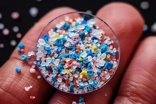
Microplastics are tiny particles of plastic that are contained in the air, plastic dust, food, fabrics, table salt, trash, and nearly every part of modern life. They can range from five millimeters to one micrometer (µm) [11]. Even smaller sizes of microplastics, called nanoplastics, pose a threat to human cells. Less than 100 nm in size, nanoplastics can cross all organs, including the placenta and blood system [11]. Microplastics of size ≤ 20 µm can enter any organ, and; ≤ 100 µm can be absorbed from the gut to the liver [11]. Scientists have discovered microplastics in many parts of the human body, including the liver, blood, and other reproductive organs, including the placenta [15].
Microplastics have multiple routes of getting into the body, which makes them a challenging threat for humans to overcome. To begin, they can be absorbed into the body by wearing clothes with fabrics containing plastic, like polyester. Although this most commonly occurs via inhalation of microplastics in the air, emerging theories also suggest that with long enough exposure to intact or open wounds, absorption of nanoplastics through the skin is possible as well. Inhalation can also occur from air pollution, specifically in areas with high carbon dioxide and dust levels.
In addition, microplastics can be consumed through foods we eat, or plastics we drink or touch, like plastic straws. Marine life also consumes a significant amount of microplastics from pollution in the ocean. Importantly for humans, this is an entry point to the food supply, as the contaminated marine life will then pass the microplastics up the food chain to humans when we eat seafood [11]. Moreover, cleaning products and cosmetics can contain a high amount of plastics that are absorbed into the skin [11]. Some estimates say that a credit card’s worth of microplastics is inhaled by an individual human every week [2].
A practical solution would be to pass the microplastics in the stool; however, the plastics do not always leave the body via waste. Sometimes, microplastics accumulate in the body over long periods of time and absorb into the intestines, bloodstream, and other tissues. Microplastics tend to find their way into crucial arteries and tissues due to their molecular composition.
They are made of synthetic polymers, a series of repeating monomers. The monomers in microplastics are made up of carbon and hydrogen atoms and occasionally have oxygen, nitrogen, chlorine, or sulfur atoms inside [3]. Some of the main components of microplastics are their polymer chains because, like polyethylene, they contain monomers like (–CH₂–CH₂–)ₙ [3]. Also, plastics usually contain additives to enhance their usual properties, but they also have harmful effects on humans. For example, phthalates, which make polyethylene flexible, negatively impact reproductive signals, while colorants are not chemically bonded to the polymer, and thus escape into the environment [3]. Most importantly, microplastics are mostly hydrophobic, which means they repel against water. This causes them to bind with oily substances and bioaccumulate in human tissues [3].
Female Reproductive System
The reproductive system is a highly complex system requiring the coordination between several organ systems and the endocrine system to ensure the human body is an adequate environment for fetal development. The hypothalamic-pituitary-gonadal axis, located between the brain and reproductive organs helps to control ovulation and coordinate reproductive behavior [8].
First, a primary signal called the GnRH (gonadotropin-releasing hormone) is produced by the hypothalamic neurons, which stimulates the pituitary gland to release two important hormones: FSH (follicle–a fluid filled sac in the ovary that contains the immature egg–stimulating hormone) and LH (luteinizing hormone) [8]. These hormones lead to ovarian growth, egg maturation, and preparation of the uterine lining for pregnancy [8]. As the follicles grow, they start to make a form of estrogen known as estradiol, which will ultimately slow down the production of GnRH and FSH [8]. Once there is an adequate amount of estradiol, the GnRH and FSH will burst and surge, leading to ovulation. These reproductive hormones, such as GnRH, regulate the proper timing of a woman’s reproductive cycle [8].
However, foreign chemicals, microplastics, and agents can interfere with hormonal signals, either blocking or mimicking them. This disruption can cause infertility, irregular menstrual cycles, and complications in fetal development, since hormones are key to regulating and protecting the growth of vital organs like the baby’s brain and heart [8].
The placenta forms in a woman during pregnancy. The placenta is crucial for fetal development as it connects the fetal and maternal circulations via the umbilical cord. It supports the baby’s growth and development by providing nutrition and removing waste from the baby’s blood. In addition, the organ plays a major role in immunity because it helps the fetus identify self versus non-self cells and antigens. The placenta is located on the wall of the uterus lining and usually on the top, side, and sometimes even the lower area. When the placenta is too low, it raises a risk known as placenta previa, which is caused when the organ covers the cervical opening, and it can develop this way if microplastics were to block and change growth signaling for the placenta [14].
Microplastics in Female Reproduction
Microplastics enter the human placenta through many of the same pathways they use to accumulate in other tissues. First, they can be introduced through food consumption or inhalation [2]. Then, particles are absorbed through the gut and travel into the bloodstream, where they find their way into the placenta during pregnancy.
On a molecular level, after entering the body, their hydrophobic polymer chains prevent normal decomposition [2]. This means microplastics can proceed and bind to other toxins such as heavy metals, which can enhance the harmful effects in living organisms. Once inside the body, the microplastics can cross membranes such as those in the gut, like the M-cells in the intestinal lining, through the cellular process of endocytosis, which can take in foreign particles [2]. From there, they can enter the lymphatic system and/or the bloodstream [2].
Another pathway for microplastics is that sometimes they can bypass the digestive system completely through cells or between cells transport, which is also known as trans-cellular and paracellular transport [2]. Once in the bloodstream, microplastics can circulate to any part of the body, including the placenta. While the placenta does have a layer to protect it from harmful substances called a syncytiotrophoblast layer, nanoplastics can bypass this layer through endocytosis or passive diffusion through functional surfaces coated with proteins [2].
Once inside, the microplastics may interact with intracellular structures like the mitochondria, which can affect energy production, the endoplasmic reticulum, and as a result impact protein synthesis and lysosomes, ultimately leading to cell damage [2]. Studies show high levels of microplastics in human placental tissue:
In a 2024 study led by Dr. Matthew Campen and colleagues, microplastics were found in all 64 placentas studied, with amounts ranging from 6.5 to 790 micrograms per gram of tissue. Moreover, it was found that 54% of the plastic was polyethylene, the plastic that makes up plastic bags and bottles, with polyvinyl chloride and nylon being 10%, and the rest being nine other polymers [13]. This suggests that a majority of the placental microplastics are likely inhaled due to direct contact with the plastics on our mouth, nose, hands, etc.
Another study showed that 10.9% of all microplastics found in a human body were in the placenta, demonstrating how common microplastic exposure is during human development [5]. Thus, microplastics can enter the developing fetus through the placenta [13]. Multiple international studies have found microplastics within the placenta and neonatal samples, suggesting a widespread exposure of microplastics globally [4]. Between 2021 and 2023, seven studies were conducted in four countries, which showed a high percentage of microplastics in the placental tissue.
In 2021, an Italian study identified microplastics in four out of six placentas from vaginal births using light microscopy and Raman microspectroscopy [9]. In another Italian study, all ten placentas (from both vaginal and Cesarean section births) contained microplastics [9]. Electron microscopy revealed cellular damage, although the association with microplastics was not definitive [9]. Importantly, higher microplastics and polymer levels were linked to greater water consumption and frequent use of certain personal care products [9].
In 2022, an Iranian study detected microplastics in 13/13 placentas from the intrauterine growth restriction (IUGR) group and only 4/30 in the normal group [9]. This study implied that microplastic exposure may affect fetal development and normal growth. More studies also showed the presence of microplastics in cord blood samples [4]. However, only a few were tested since there is no commercially available test to find microplastics in placentas. These studies demonstrate that, as reproduction continues, this cycle could lead to a growing buildup of microplastics in future offspring and a possibility of new illnesses that will go unnoticed.
Placental microplastics affect reproduction and early fetal development. Fetal development begins from the first stage of pregnancy, often before many women realize they are pregnant [19]. There are three stages of fetal development: germinal, embryonic, and fetal [19]. The germinal stage is where the sperm and egg combine to form the zygote [19]. From there, the zygote turns into a blastocyst, where it is implanted into the uterus [19]. Next is the embryonic stage, usually from around the third week of pregnancy to the eighth week [19]. During this stage, the blastocyst becomes an embryo as the baby develops human characteristics such as organs [19]. At weeks five to six, the heart is recognized in the baby, and little arm and leg stubs are also discoverable [19]. Finally, the fetal stage begins around the ninth week and lasts until birth. During the fetal stage, the baby develops its primary sex characteristics that officially turn the embryo into a fetus. The fetus also grows hair and fingernails at this time and can start to move [19].
Microplastics can affect fetal development in several ways. Ultimately, babies are born pre-polluted [12].
“If we are seeing effects on placentas, then all mammalian life on this planet could be impacted,” says Dr. Matthew Campen, Regents’ Professor, UNM Department of Pharmaceutical Sciences.
Once the microplastics and nanoplastics enter cells, including both germ and somatic cells, they can cause oxidative damage, which can lead to DNA damage and cell death [16].
Microplastics can lead to cell death through pyroptosis [16], a highly inflammatory form of lytic programmed cell death caused by microbial infection [17]. When microplastics are detected, there is trafficking of immune cells like natural killer, T cells, and uterine dendritic cells to extinguish them as they are detected as non-self [16]. In mouse models, placental microplastics were shown to reduce the number of live births, alter the sex ratio of offspring, and cause fetal growth restriction, all effects that have also been observed in humans.
If one of these effects is already seen in humans, it raises the possibility that the others could follow. Since microplastics are present in human tissues, the outcomes seen in animal models like hormonal disruption, reduced sperm count and viability, decreased egg quality, neurophysiological and cognitive deficits, and disrupted embryonic development, [1] could also emerge in humans.
Furthermore, microplastics can change the gut microbiome and hormonal signaling, which can directly impact normal physiology and alter the signals sent between the uterus and embryo [1]. They do this by changing the balance and composition of the gut, which can lead to dysbiosis, an imbalance of the gut bacteria [10]. Some changes to the delicate gut microbiome could cause a condition called leaky gut, which shifts the previously semi-permeable membrane into a hyperpermeable one [10]. Emerging research demonstrates increasing rates of infertility, with scientists implicating environmental exposures, including microplastics.
Microplastics may also affect the endocrine system, which leads to neurodevelopmental issues in the offspring [1]. Another feature of abnormal pregnancies can be high blood pressure in mothers (like preeclampsia), which can result in organ failure and severe problems in the mother [1]. The endocrine system is the hormone-regulating system in your body that directly involves the glands of the gonads (ovaries and testes). Microplastics can interfere with the production of these hormones due to the additive factors the polymers carry, like Bisphenol A (BPA), which is used to harden the plastic [1].
These chemicals are known as endocrine-disrupting chemicals. In addition to this, it can directly bind to the hormone receptors and block normal signaling [1]. Such effects can change gene expression, cause hormone-related cancers, and most importantly, impact fetal endocrine function and development, including lower birth weight and reproductive disorders [1]. Ovarian cysts—fluid-filled sacs that develop on or in the ovaries—can also be caused by microplastics in the reproductive system [15]. When a hormone signal is out of balance, it can trigger the egg not to be released, which can persist to form a cyst [15]. Although this is still being researched by scientists today, there has been a direct correlation in mice, suggesting microplastics disrupt ovarian follicle development.
While the immediate effects of microplastics in placentas are concerning, there are other long-term concerns, such as a generational impact, that raise a sense of urgency to the issue. First, microplastics do not disappear once a person dies [6]. The synthetic particles of microplastics resist biodegradation when the body is buried or even cremated [6]. This means it can reenter the ecosystem and harm other organisms [6]. On the other hand, microplastics are also being passed from generation to generation through parental gametes and the placenta. Microplastics can lead to more detrimental impacts that haven’t even been discovered yet. With more and more accumulation, the body can respond in many different ways that are hard to predict. However, it can be assumed that populations with more microplastics are more likely to be infertile in the future. One can imagine a scenario in which natural selection might occur, as people with less microplastics or who are less affected by their presence will be better able to survive and reproduce.
Summary and Conclusion
Microplastics lead to hormone imbalances of estrogen and other hormones in female bodies by disrupting hormone signaling (activating and blocking), and altering reproductive organ function and development, including infant birth weight, length, and head circumference [10]. Microplastics can interfere with gene expression or epigenetic markers, which can alter the way a fetus develops [10]. They can cut gene readings short, which could lead to affecting their length or head circumference [10]. Impaired egg development and follicular growth can impair fertility and have been linked with microplastic exposure [10]. Similarities can be seen in male fertility as microplastics affect the inflammatory response, change hormone levels with their disrupting and toxic chemicals, and cause cellular damage to the development of the gametes [5]. Overall, the effects of microplastics on reproductive systems have grave consequences, with evidence suggesting infertility in humans.
In addition to understanding the effects of microplastics on human health and reproduction, scientists are working to rid the body of microplastics. By studying plastic-eating microorganisms, they can examine the enzymes they have that allow them to process microplastics naturally [10]. Additionally, as there is increasing understanding of methods of exposure, such as inhalation or absorption, [10], there are ways to reduce the chance of microplastic exposure to your body. For example, humans face the biggest possibility of exposure from food. Fish is a great source of nutrients and protein, however, it is extremely crucial to know that fish carry large quantities of microplastics ingested in the ocean. By ensuring trash and plastics do not end up in aquatic ecosystems, humans can reduce the chance of microplastics entering the food chain. Scientists are also advocating for the elimination of single-use plastic and finding a more sustainable way to save the human population and the environment.
A Simple Technique for Studying the Interaction of Polypropylene-Based Microplastics with Adherent Mammalian Cells Using a Holder. Feb. 2025, research.ebsco.com/c/3uzxq3/search/details/hul46wuiu5?isDashboardExpanded=true&limiters=FT1%3AY&q=DE%20%22MICROPLASTICS%22.
Chemical Analysis of Microplastics and Nanoplastics: Challenges, Advanced Methods, and Perspectives. 26 Aug. 2021, pubs.acs.org/doi/10.1021/acs.chemrev.1c00178.
Cleveland Clinic. “Fetal Development.” Cleveland Clinic, 19 Mar. 2024, my.clevelandclinic.org/health/articles/7247-fetal-development-stages-of-growth.
Comparison of Microplastic Levels in Placenta and Cord Blood Samples of Pregnant Women With Fetal Growth Retardation and Healthy Pregnant Women. Kutahya Health Sciences University, 1 Apr. 2022. clinicaltrials.gov/study/NCT05070715?cond=placenta&term=microplastics&rank=1.
Exposure to Microplastics and Human Reproductive Outcomes: A Systematic Review. 29 Jan. 2024, obgyn.onlinelibrary.wiley.com/doi/10.1111/1471-0528.17756.
Haederle, Michael. “Microplastics in Every Human Placenta, New UNM Health Sciences Research Discovers.” UNM HSC Newsroom, 2024, hscnews.unm.edu/news/hsc-newsroom-post-microplastics.
Hunt K, Davies A, Fraser A, Burden C, Howell A, Buckley K, et al. Exposure to microplastics and human reproductive outcomes: A systematic review. BJOG. 2024; 131(5): 675–683. https://doi.org/10.1111/1471-0528.17756.
Leaky Gut Syndrome. Cleveland Clinic, 6 Apr. 2022, my.clevelandclinic.org/health/diseases/22724-leaky-gut-syndrome.